. 2021 Dec 16;12(12):1998.
doi: 10.3390/genes12121998.

Cell-Cycle-Dependent Chromatin Dynamics at Replication Origins

Affiliations

Cell-Cycle-Dependent Chromatin Dynamics at Replication Origins

Yulong Li et al. Genes (Basel). .

Abstract

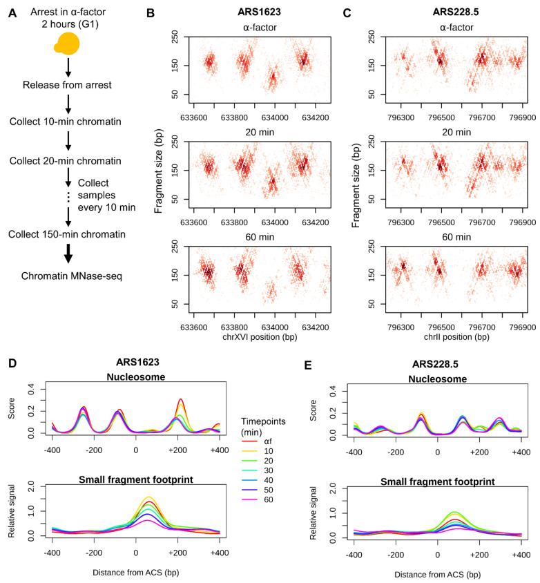
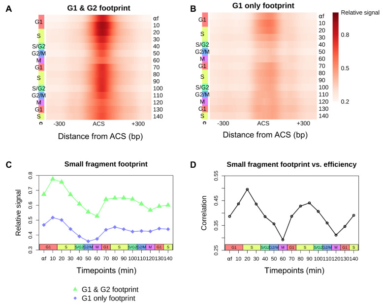
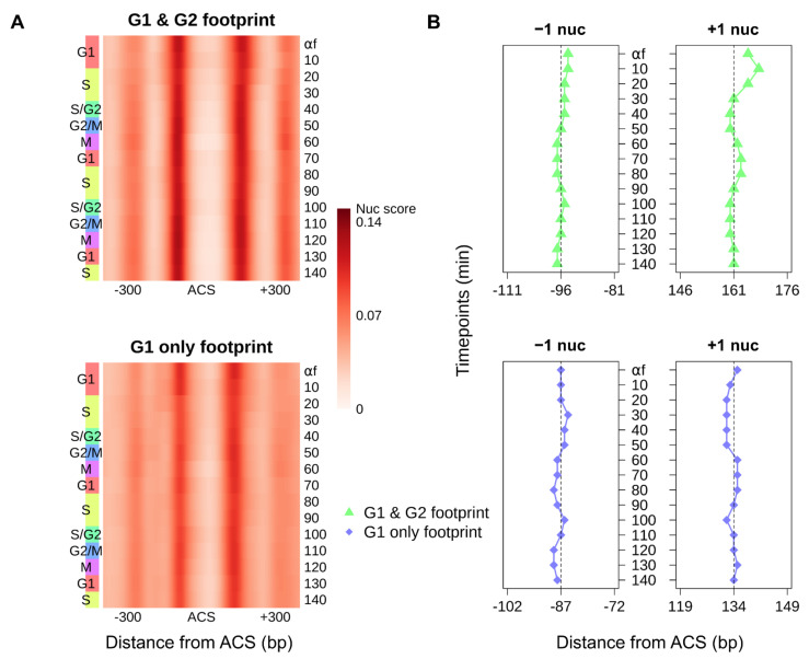
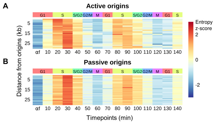
Origins of DNA replication are specified by the ordered recruitment of replication factors in a cell-cycle-dependent manner. The assembly of the pre-replicative complex in G1 and the pre-initiation complex prior to activation in S phase are well characterized; however, the interplay between the assembly of these complexes and the local chromatin environment is less well understood. To investigate the dynamic changes in chromatin organization at and surrounding replication origins, we used micrococcal nuclease (MNase) to generate genome-wide chromatin occupancy profiles of nucleosomes, transcription factors, and replication proteins through consecutive cell cycles in Saccharomyces cerevisiae. During each G1 phase of two consecutive cell cycles, we observed the downstream repositioning of the origin-proximal +1 nucleosome and an increase in protected DNA fragments spanning the ARS consensus sequence (ACS) indicative of pre-RC assembly. We also found that the strongest correlation between chromatin occupancy at the ACS and origin efficiency occurred in early S phase, consistent with the rate-limiting formation of the Cdc45-Mcm2-7-GINS (CMG) complex being a determinant of origin activity. Finally, we observed nucleosome disruption and disorganization emanating from replication origins and traveling with the elongating replication forks across the genome in S phase, likely reflecting the disassembly and assembly of chromatin ahead of and behind the replication fork, respectively. These results provide insights into cell-cycle-regulated chromatin dynamics and how they relate to the regulation of origin activity.

Keywords: DNA replication; cell cycle; chromatin; replication origins.


Conflict of interest statement

The authors declare no conflict of interest.

Figures

Figure 1
Figure 1
Profiling cell-cycle–dependent chromatin dynamics by MNase mapping. (A) Schematic of the experimental design for capturing cell-cycle–dependent chromatin dynamics. (B) Chromatin profiles at ARS1623 for select time points during the first cell cycle. The midpoints of recovered and sequenced MNase fragments are displayed. The size of each fragment is plotted as a function of its midpoint chromosomal position. (C) Chromatin profiles at ARS228.5 for select time points during the first cell cycle. (D,E) Quantification of nucleosome scores and small fragment (<120 bp) occupancy at all time points for the first cell cycle at ARS1623 and ARS228.5, respectively. The same chromosome regions as in (B,C) are shown for each ARS locus.
Figure 2
Figure 2
The cell-cycle–Dependent accumulation of small protected fragments at ACS sites correlates with origin efficiency. (A,B) Heatmaps of aggregate small fragment (<120 bp) occupancy at 264 origins with a previously described ORC-dependent footprint in both “G1 & G2” (A) and 128 origins with an ORC-dependent footprint in “G1 only” (B) plotted throughout the cell cycle [20]. All origins are oriented by the T-rich ACS strand. (C) Average small fragment occupancy ±100 bp surrounding the peak of the aggregate ORC-dependent footprint for each class of origins. (D) Spearman correlation between log2-transformed ACS-bound small fragment footprint density and activation efficiency [2] for 371 origins exhibiting an ORC-dependent footprint at each time point.
Figure 3
Figure 3
Cell-cycle–dependent nucleosome dynamics at replication origins. (A) Heatmaps of aggregate nucleosome scores for 264 origins with an ORC-dependent footprint in both “G1 & G2” (top panel) and 128 origins with an ORC-dependent footprint in “G1 only” (bottom panel) throughout the cell cycle [20]. All origins are oriented by the T-rich ACS strand. (B) Dyad positions of the aggregate −1 and +1 nucleosomes relative to the ACS for each cell-cycle time point and for each class of origins.
Figure 4
Figure 4
Nucleosome disruption at the replication fork. (A,B) Heatmaps representing the average nucleosome entropy at each time point in 1 kb windows for 30 kb surrounding the top 20% (n = 69) most efficient (active) origins (A) and the bottom 20% (n = 69) least efficient (passive) origins (B), among origins exhibiting an ORC-dependent footprint [2]. Each row represents a genomic window and rows are ordered by the distance of that window from the nearest origin. Nucleosome entropy is standardized into z-scores across each row.

References

    1. Lin Y.C., Prasanth S.G. Replication initiation: Implications in genome integrity. DNA Repair. 2021;103:103131. doi: 10.1016/j.dnarep.2021.103131. - DOI - PMC - PubMed
    1. McGuffee S.R., Smith D.J., Whitehouse I. Quantitative, Genome-Wide Analysis of Eukaryotic Replication Initiation and Termination. Mol. Cell. 2013;50:123–135. doi: 10.1016/j.molcel.2013.03.004. - DOI - PMC - PubMed
    1. Aparicio O.M. Location, location, location: It’s all in the timing for replication origins. Genes Dev. 2013;27:117–128. doi: 10.1101/gad.209999.112. - DOI - PMC - PubMed
    1. Wyrick J.J., Aparicio J.G., Chen T., Barnett J.D., Jennings E.G., Young R.A., Bell S.P., Aparicio O.M. Genome-Wide Distribution of ORC and MCM Proteins in S. cerevisiae: High-Resolution Mapping of Replication Origins. Science. 2001;294:2357–2360. doi: 10.1126/science.1066101. - DOI - PubMed
    1. Aparicio O.M., Weinstein D.M., Bell S.P. Components and Dynamics of DNA Replication Complexes in S. cerevisiae: Redistribution of MCM Proteins and Cdc45p during S Phase. Cell. 1997;91:59–69. doi: 10.1016/S0092-8674(01)80009-X. - DOI - PubMed

Publication types