. 2021 Dec 20;7(12):1097.
doi: 10.3390/jof7121097.

Phylogenetic Reassessment, Taxonomy, and Biogeography of Codinaea and Similar Fungi

Affiliations

Phylogenetic Reassessment, Taxonomy, and Biogeography of Codinaea and Similar Fungi

Martina Réblová et al. J Fungi (Basel). .

Abstract

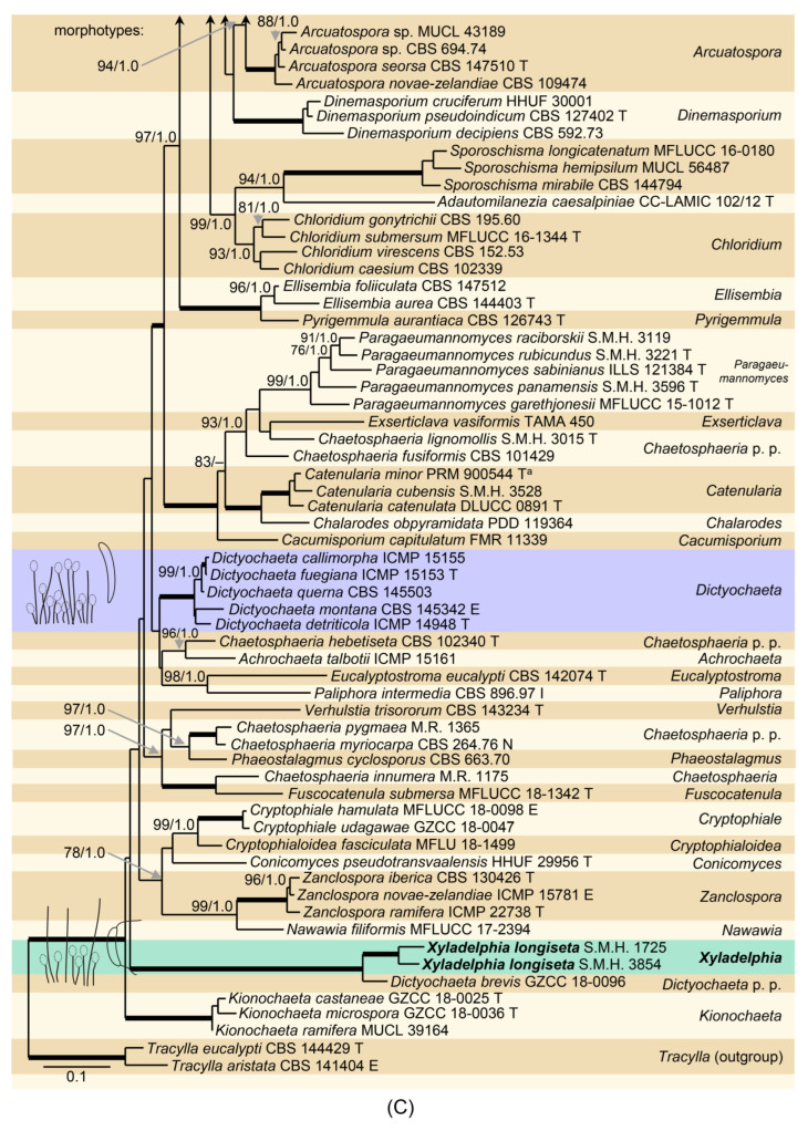
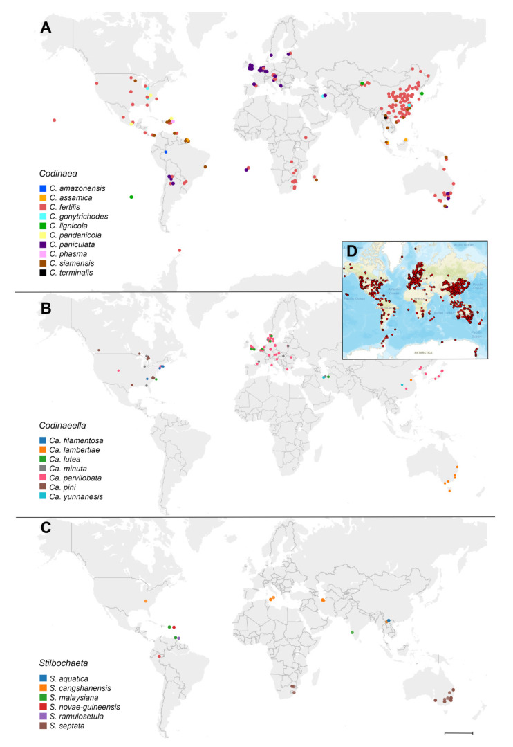
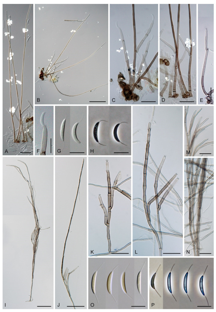
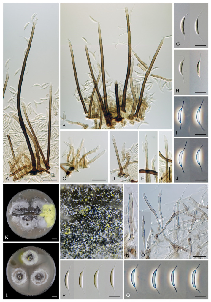
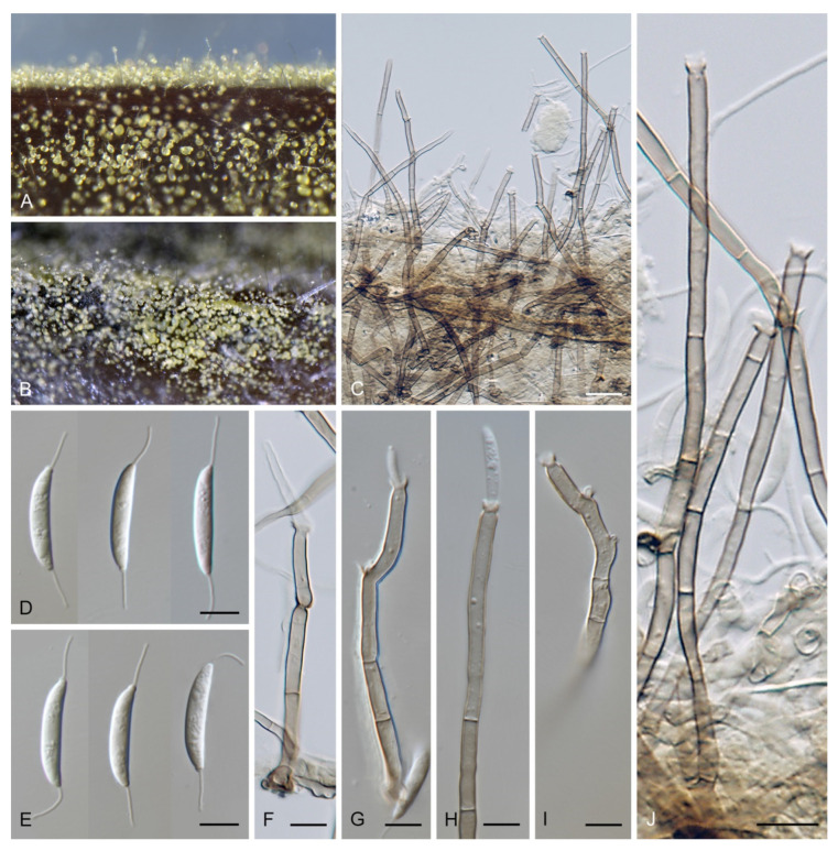
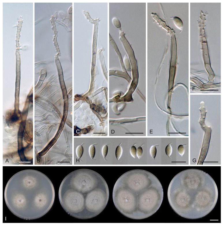
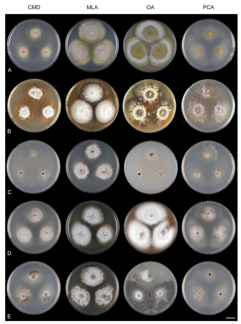
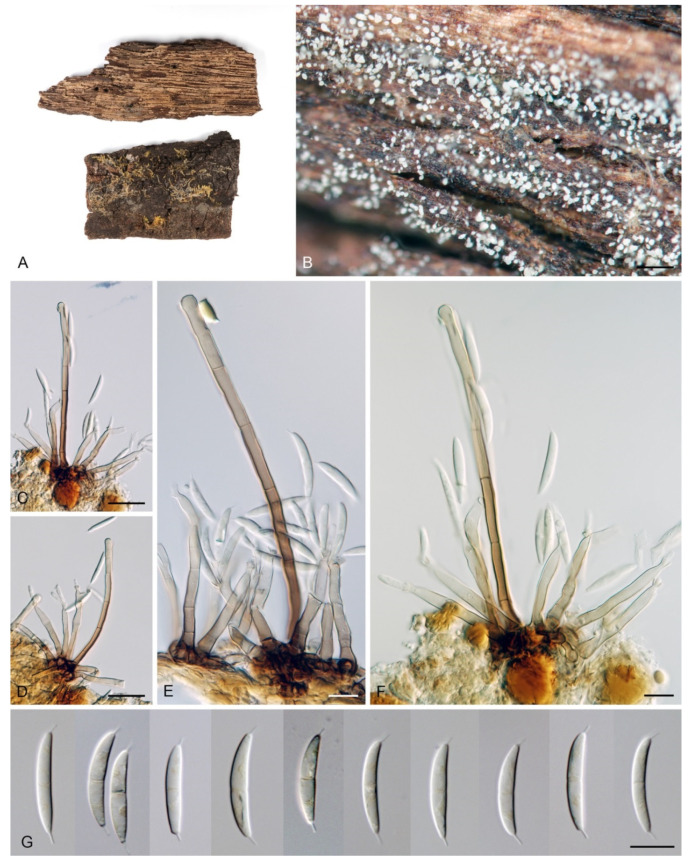
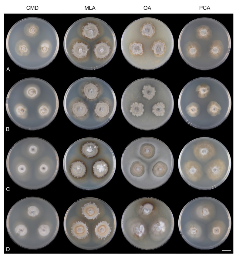
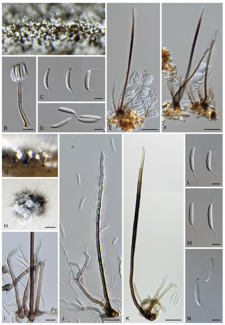
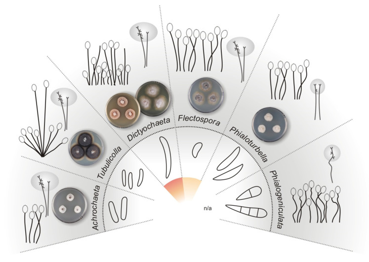
The genus Codinaea is a phialidic, dematiaceous hyphomycete known for its intriguing morphology and turbulent taxonomic history. This polyphasic study represents a new, comprehensive view on the taxonomy, systematics, and biogeography of Codinaea and its relatives. Phylogenetic analyses of three nuclear loci confirmed that Codinaea is polyphyletic. The generic concept was emended; it includes four morphotypes that contribute to its morphological complexity. Ancestral inference showed that the evolution of some traits is correlated and that these traits previously used to delimit taxa at the generic level occur in species that were shown to be congeneric. Five lineages of Codinaea-like fungi were recognized and introduced as new genera: Codinaeella, Nimesporella, Stilbochaeta, Tainosphaeriella, and Xyladelphia. Dual DNA barcoding facilitated identification at the species level. Codinaea and its segregates thrive on decaying plants, rarely occurring as endophytes or plant pathogens. Environmental ITS sequences indicate that they are common in bulk soil. The geographic distribution found using GlobalFungi database was consistent with known data. Most species are distributed in either the Holarctic realm or tropical geographic regions. The ancestral climatic zone was temperate, followed by transitions to the tropics; these fungi evolved primarily in Eurasia and Americas, with subsequent transitions to Africa and Australasia.

Keywords: 37 new taxa; GlobalFungi; ancestral inference; appendages; barcodes; molecular systematics; morphology; phialidic conidiogenesis.


Conflict of interest statement

The authors declare no conflict of interest. The funders had no role in the design of the study; in the collection, analyses, or interpretation of data; in the writing of the manuscript, or in the decision to publish the results.

Figures

Figure 1
Figure 1
Illustration of a seta, conidiophores, and conidia of Codinaea aristata (holotype, Maire 1937).
Figure 2
Figure 2
(A) Phylogenetic tree based on the combined ITS-28S-tef1-α rDNA sequences constructed by maximum likelihood (RAxML) of members of the Chaetosphaeriaceae. Species names given in bold and placed in green boxes are taxonomic novelties. Each novelty is accompanied by a respective morphotype(s); their distribution within the genus is indicated by circles of different colors. Codinaea: morphotypes C1 (dark violet), C2 (fuchsia), C3 (lila), C4 (pink); Codinaeella: morphotypes CA1 (yellow), CA2 (burgundy). The clade containing Dictyochaeta s. str. is placed in a violet box. Abbreviations T, E, I, N, and P indicate ex-type, ex-epitype, ex-isotype, ex-neotype and ex-paratype strains; letters in the upper case after a species name indicate: (a) holotype of Chaetosphaeria trianguloconidia, (b) ex-type strain of Codinaea acaciae, (c) ex-type strain of Chaetosphaeria ciliata, (d) ex-type strain of Dictyochaeta curvispora (contamination), (e) ex-type strain of Tainosphaeria obclavata. Thickened branches indicate branch support with ML BS = 100% and PP values = 1.0. Branch support of nodes ≥ 75% ML and ≥0.95 PP is indicated above or below branches. (B,C) Phylogenetic trees based on the combined ITS-28S-tef1-α rDNA sequences of the Chaetosphaeriaceae (continued). For legend refer to (A). Abbreviation: p.p. after a genus name (pro parte).
Figure 2
Figure 2
(A) Phylogenetic tree based on the combined ITS-28S-tef1-α rDNA sequences constructed by maximum likelihood (RAxML) of members of the Chaetosphaeriaceae. Species names given in bold and placed in green boxes are taxonomic novelties. Each novelty is accompanied by a respective morphotype(s); their distribution within the genus is indicated by circles of different colors. Codinaea: morphotypes C1 (dark violet), C2 (fuchsia), C3 (lila), C4 (pink); Codinaeella: morphotypes CA1 (yellow), CA2 (burgundy). The clade containing Dictyochaeta s. str. is placed in a violet box. Abbreviations T, E, I, N, and P indicate ex-type, ex-epitype, ex-isotype, ex-neotype and ex-paratype strains; letters in the upper case after a species name indicate: (a) holotype of Chaetosphaeria trianguloconidia, (b) ex-type strain of Codinaea acaciae, (c) ex-type strain of Chaetosphaeria ciliata, (d) ex-type strain of Dictyochaeta curvispora (contamination), (e) ex-type strain of Tainosphaeria obclavata. Thickened branches indicate branch support with ML BS = 100% and PP values = 1.0. Branch support of nodes ≥ 75% ML and ≥0.95 PP is indicated above or below branches. (B,C) Phylogenetic trees based on the combined ITS-28S-tef1-α rDNA sequences of the Chaetosphaeriaceae (continued). For legend refer to (A). Abbreviation: p.p. after a genus name (pro parte).
Figure 3
Figure 3
The frequency distribution graphs of the Kimura 2-parameter distances of ITS and tef1-α DNA sequences (barcoding gaps). (A) Codinaea; (B) Codinaeella; (C) Stilbochaeta. The intraspecific distances are shown as blue bars and interspecific distances as grey bars. The bin size of 0.001 and 0.0005 (only ITS, Codinaeella) were used.
Figure 4
Figure 4
ITS2 secondary structure of Codinaea assamica CBS 139907 and 5.8S-28S rRNA gene hybridization (proximal stem region) (GenBank accessions ITS: OL654077, 28S: OL654134). ITS2 helices are numbered I–III. All substitutions recorded among members of Codinaea are mapped on the 2D model. Identified substitutions are color-coded: CBC (green), hCBC (blue), and non-CBC (red). Parts of the text in green frames refer to CBCs, parts of the text in blue and red refer to hCBCs and non-CBCs, respectively. Hairpin loops and other parts of the ITS2 molecule highlighted with grey color represent regions with a variable number of nucleotides or sequence variation; positions highlighted with a pink color represent single mutations.
Figure 5
Figure 5
ITS2 secondary structure of Codinaeella and Stilbochaeta and 5.8S-28S rRNA gene hybridization (proximal stem region). (A) Codinaeella minuta CBS 146620 (GenBank accessions ITS: OL654095, 28S: OL654152) (B) Stilbochaeta malaysiana IMI 312436 (GenBank accessions ITS: OL654121, 28S: OL654178). Details as in Figure 4.
Figure 6
Figure 6
Reconstruction of ancestral states and areas in Rasp v.4.2 using phylogenetic trees calculated in MrBayes v.3.2.7. (AD) Reconstruction of morphological characters using Multistate function in Bayestraits (E,F) ancestral areas reconstruction of species distribution using Bayesian Binary MCMC (BBM) method.
Figure 7
Figure 7
Geographical distribution of (A) Codinaea, (B) Codinaeella, and (C) Stilbochaeta species based on the GlobalFungi database. See Table S2 for primary data. Species are differentiated by colors. (D) The circles represent smaller geographical areas with one or more observations of the particular species. Some of the smaller areas contained observations of multiple species (e.g., Caspian coast, Puerto Rico); circles were manually dispersed over the actual collection site. Scale bar: 2000 km.
Figure 8
Figure 8
Illustrations of morphotypes based on the arrangement of setae, conidiophores and conidiogenous cells. (A) Codinaea morphotypes C1–C4 (B) Codinaeella morphotypes CA1–CA2 (C) Chloridium morphotypes CH1–CH3 (D) Menispora morphotypes M1–M4.
Figure 9
Figure 9
Selected morphotypes of Menispora and Chloridium. (A) M. ciliata morphotype M2 (CBS 122131); (B) M. tortuosa morphotype M3 (CBS 117552) (C,D) M. uncinata morphotype M4 (ICMP 18253) (E) M. britannica morphotype M1 (PRA-20985) (F) Chl. virescens morphotype CH1 (M.R. 3886) (G) Chl. caesium morphotype CH3 (M.R. 1883). Scale bars: (A,B,D,E) 20 μm; (C) = 100 μm; (F,G) 50 μm.
Figure 10
Figure 10
Codinaea amazonensis (MUCL 41171). (AC) Conidiophores (DG) conidiogenous cells (HJ) conidia. Images: (AJ) on MLA after 4 weeks. Scale bars: (AC) 20 μm; (DJ) 10 μm.
Figure 11
Figure 11
Colony morphology of Codinaea spp. after 4 weeks. (A) C. amazonensis MUCL 41171 (B) C. gonytrichodes CBS 593.93 (C) C. phasma CBS 147516 ex-type. Scale bar: (AC) 1 cm.
Figure 12
Figure 12
Codinaea assamica (CBS 139907 ex-type of C. acaciae). (AE) Setae with conidiophores (FH) conidia (I) apical part of the setae. Images: (AI) on pine needles on SNA after 4 weeks. Scale bars: (A,D) 25 μm; (B) 50 μm; (C,E) 20 μm; (FI) 10 μm.
Figure 13
Figure 13
Colony morphology of Codinaea spp. after 4 weeks. (A) C. assamica CBS 139907 ex-type of C. acaciae (B) C. fertilis IMI 233824 (C) C. fertilis MUCL 15427 (D) C. dwaya CBS 261.77 ex-type (E) C. siamensis CBS 194.96. Scale bar: (AC) 1 cm.
Figure 14
Figure 14
Codinaea dwaya (CBS 261.77 ex-type). (AG) Conidiophores (HJ) conidia (KO) microconidia. Images: (AO) on stems of U. dioica on CMA after 8 weeks. Scale bars: (AG) 20 μm; (HO) 10 μm.
Figure 15
Figure 15
Codinaea fertilis (DAOM 93548c isotype). (A) Substrate with colonies (B) colony on a dead leaf (C,D) conidiophores (E,F) apices of setae with phialidic apertures (G,H) conidia. Images: (AH) from nature. Scale bars: (B) 500 μm; (C) 25 μm; (D) 20 μm; (EH) 10 μm.
Figure 16
Figure 16
Codinaea fertilis (IMI 233824). (AD) Setae with conidiophores (E) conidia (F,G) conidiogenous cells at the tip of the setae. Images: (AG) on PCA after 6 weeks. Scale bars: (A,B) = 50 μm; (C,D) 20 μm; (EG) 10 μm.
Figure 17
Figure 17
Codinaea gonytrichodes (CBS 593.93). (AD) Conidiophores (E) conidia (FH) conidiogenous cells arising from collar hyphae (I,J) conidiogenous cells at the tip of the conidiophore. Images: (AJ) on OA after 8 weeks. Scale bars: (A,B) 20 μm; (C,D) 50 μm; (EJ) 10 μm.
Figure 18
Figure 18
Colony morphology of Codinaea paniculata after 4 weeks. (A) CBS 145098 ex-type (B) CBS 127692 (C) MUCL 34876. Scale bar: (AC) 1 cm.
Figure 19
Figure 19
Codinaea phasma (CBS 147516 ex-type). (A,B,I) Colonies (C,D) setae (E,F) setae with conidiophores (G) conidiogenous cell (H,M,N) conidia (J,K) apices of the conidiogenous cells, conidiogenous locus slightly protruding above the collarette (L) conidium, from the top view (OQ) conidiophores. Images: (AH) from nature (IQ) on stems of U. dioica on CMA after 4 weeks. Scale bars: (CF) 25 μm; (G,H,JQ) 5 μm.
Figure 20
Figure 20
Codinaea siamensis (CBS 194.96). (A,B) Setae with conidiophores (C) conidiophores (DF) conidiogenous cells with collarettes (G,H) conidia. Images: (AH) on MLA after 6 weeks. Scale bars: (A) 50 μm; (B,C) 20 μm; (DH) 10 μm.
Figure 21
Figure 21
Codinaeella filamentosa (CBS 147265). (AE,IN) Conidiophores (F) conidiogenous cell (G,H,O,P) conidia. Images: (AH) from nature (IP) on MLA after 4–8 weeks. Scale bars: (A) 20 μm; (B,I,J) 50 μm; (CE,KN) 20 μm; (FH,O,P) 10 μm.
Figure 22
Figure 22
Colony morphology of Codinaeella spp. after 4 weeks. (A) Ca. filamentosa CBS 147265 (B) Ca. lambertiae CBS 143419 ex-type (C) Ca. mimusopis CBS 143435 ex-type (D) Ca. pini CBS 138866 ex-type. Scale bar: (AD) 1 cm.
Figure 23
Figure 23
Codinaeella lamberiae (CBS 143419 ex-type). (AD) Conidiophores (E,F) conidia. Images: (AD) on OA after 4 weeks. Scale bars: (A) 50 μm; (BF) 10 μm.
Figure 24
Figure 24
Codinaeella lutea. (AF,N,O) Conidiophores (GJ,P,Q) conidia (KM) colonies. Images: (AJ) from nature (K,MQ) on stems of U. dioica on CMA after 8 weeks (L) on OA after 4 weeks (AL) from CBS 146618 ex-type (MQ) from CBS 624.77. Scale bars: (AF,O) 20 μm; (K,L) 1 cm; (N,P,Q) 10 μm.
Figure 25
Figure 25
Colony morphology of Codinaeella lutea after 4 weeks. (A) CBS 146618 ex-type (B) CBS 624.77 (C) ICMP 14613 (D) ICMP 15544. Scale bar: (AD) 1 cm.
Figure 26
Figure 26
Codinaeella mimusopis (CBS 143435 ex-type). (A,B) Colonies (C,FJ) conidiophores (D,E) conidia. Images: (A) on a pine needle on SNA after 8 weeks (B) on a stem of U. dioica on CMA after 8 weeks. Scale bars: (C,FI) 20 μm; (D,E) 5 μm; (J) 10 μm.
Figure 27
Figure 27
Codinaeella minuta. (AD,IM) Conidiophores (E,N) conidia (FH) colonies. Images: (AE) from nature (FH) on MLA after 4 weeks (IN) on a stem of U. dioica on CMA after 4–8 week (AE) from CBS 145099 (F) from CBS 146619 (G,H,L,M) from DAOM 148141 (IK,N) from CBS 146619. Scale bars: (AE,JN) 10 μm; (FH) 1 cm; (I) 20 μm.
Figure 28
Figure 28
Colony morphology of Codinaeella minuta after 4 weeks. (A) CBS 966.69 (B) CBS 146621 (C) CBS 115959 (D) DAOM 148141 (E) ATCC 20960. Scale bar: (AE) 1 cm.
Figure 29
Figure 29
Codinaeella parvilobata (CBS 144536 ex-type). (AG) Conidiophores (H,I) conidia (JN) conidiophores (O,P) conidia. Images: (AI) from natural substrate (JP) on MLA after 4 weeks. Scale bars: (AD) 20 μm; (EP) 10 μm.
Figure 30
Figure 30
Colony morphology of Codinaeella parvilobata after 4 weeks. (A) CBS 144536 ex-type (B) CBS 144792 (C) CBS 144658 (D) MUCL 28054. Scale bar: (AD) 1 cm.
Figure 31
Figure 31
Codinaeella pini (CBS 138866 ex-type). (A) Colony (B,C) conidia (D) conidiogenous cells (EG) conidiophores. Images: (AI) on a stem of U. dioica on CMA after 8 weeks. Scale bars: (BD) 10 μm; (EG) 20 μm.
Figure 32
Figure 32
Nimesporella capillacea (IMI 358908 ex-type). (AG) Conidiophores (H) conidia (I) colonies on CMD, MLA, OA and PCA after 4 week (from left to right). Images: (AH) on PCA after 4–6 weeks. Scale bars: (AH) 10 μm; (I) 1 cm.
Figure 33
Figure 33
Stilbochaeta aquatica (CBS 114070, ex-type strain of Dictyochaeta curvispora). (A,B) Setae with conidiophores (CE) conidiophores (FH) conidia. Images: (AH) on a stem of U. dioica on CMA after 6–8 weeks. Scale bars: (A,B) 25 μm (CH) 10 μm.
Figure 34
Figure 34
Colony morphology of Stilbochaeta spp. after 4 weeks. (A) S. aquatica CBS 114070 (B) S. malaysiana IMI 312436 ex-type (C) S. ramulosetula IMI 313452 ex-epitype (D) S. septata CBS 143386 ex-epitype (E) S. septata CBS 146716. Scale bar: (AE) 1 cm.
Figure 35
Figure 35
Stilbochaeta brevisetula (DAOM 96420a isotype). (A) Substrate with colonies (B) colony on wood (CF) setae and conidiophores (G) conidia. Images: (AG) from nature. Scale bars: (B) 500 μm; (C,D) 25 μm; (EG) 10 μm.
Figure 36
Figure 36
Stilbochaeta brevisetula. (AD) Ascomata (E) setae on the ascomal wall (F) vertical section of the ascomal wall (G,H,OR) setae and conidiophores (I) asci with ascospores (JN) conidia. Images: (AK) from nature (LR) on MLA after 6–8 weeks (A,L) from ICMP 22548 (B–E,G,H,J,K) from ICMP 22549 ex-epitype (F,I,M,N,R) from ICMP 22551 (OQ) from ICMP 22552. Scale bars: (AD) 250 μm; (EH,OR) 20 μm; (IN) 10 μm.
Figure 37
Figure 37
Colony morphology of Stilbochaeta brevisetula after 4 weeks. (A) ICMP 22548 (B) ICMP 22549 ex-epitype (C) ICMP 22551 (D) ICMP 22552. Scale bar: (AD) 1 cm.
Figure 38
Figure 38
Stilbochaeta malaysiana (IMI 312436 ex-type). (A,B) Setae and conidiophores (C,D) conidiophores with conidiogenous cells, in detail (E,F) colonies (GJ) tip of the conidiogenous cells with collarettes (K,L) conidia. Images: (AD,GL) on CMD after 4–6 weeks (E) on MLA after 4 weeks (F) on OA after 4 weeks. Scale bars: (A,B) 25 μm; (C,D) 20 μm; (E,F) 1 cm; (GL) 10 μm.
Figure 39
Figure 39
Stilbochaeta novae-guineensis. (A,G) Colonies (B,I) conidiophores (C,D,LN) conidia (E,F,J,K) setae with conidiophores (H) setae and conidiophores arranged in a sporodochium-like. Images: (AF) from nature (G,J,LN) on a stem of U. dioica on CMA after 4 weeks (H,I,K) on OA after 8 weeks (A,G,J,LN) from CBS 147515 (H,I,K) from CBS 147517. Scale bars: (B,I) 10 μm; (C,D,LN) 5 μm; (E,F) 25 μm; (H) 500 μm; (J,K) 20 μm.
Figure 40
Figure 40
Colony morphology of Stilbochaeta novae-guineensis after 4 weeks. (A) CBS 147515 (B) CBS 147517. Scale bar: (A,B) 1 cm.
Figure 41
Figure 41
Stilbochaeta ramulosetula (IMI 313452 ex-epitype). (A) Seta with conidiophores (B,C) conidiophores (D) conidia. Images: (AC) on CMD after 5 months. Scale bars: (A) 25 μm; (B,C) 20 μm; (D) 10 μm.
Figure 42
Figure 42
Stilbochaeta septata (CBS 146716). (AE) Setae with conidiophores (FI) conidia. Images: (A,B,F,G) from nature (C,H,I) on PCA after 4 weeks (D,E) on OA after 4 weeks. Scale bars: (AC) 20 μm; (DI) 10 μm.
Figure 43
Figure 43
Tainosphaeria parva (DAOM 93565d isotype). (A) Substrate with colonies (BE) conidiophores (F,G) tips of the conidiogenous cells (H) conidia. Images: (AH) from nature. Scale bars: (B) 500 μm; (CH) 10 μm.
Figure 44
Figure 44
Tainosphaeria simplex (DAOM 96020g isotype). (A) Substrate with colonies (BG) conidiophores (H) colony on wood (I) conidia. Images: (AH) from nature. Scale bars: (BG,I) 10 μm; (H) 500 μm.
Figure 45
Figure 45
Tainosphaeria vulgaris. (AE,O,P) Conidiophores (O) associated with conidiophores of Helicoma sp. (F,G) tips of the phialides (HJ,M,N) conidia (K) substrate with colonies (L) colony on wood, in detail. Images: (AD,H,I,KP) from nature (EG,J) on PCA after 4 weeks; (AJ) from PDD 119682 (KP) from DAOM 97315b isotype. Scale bars: (AE,O,P) 20 μm; (F,G) 5 μm; (HJ,M,N) 10 μm.
Figure 46
Figure 46
Morphotypes of Codinaea and similar genera with setulate conidia. Conidia and pigments formed in vitro are shown in the central (white) part of the wheel. The arrangement of conidiophores with setae, if present, of each genus, along with the morphology of the phialide (i.e., with a terminal or lateral opening) and colony characters are shown in the outer (grey) part of the wheel. Pigments diffusing in MLA and OA are shown by their respective colors and colony images; if no pigment is formed, the place at the centre is blank, n/a indicates that this information is not available.
Figure 47
Figure 47
Morphotypes of Dictyochaeta and similar genera with non-setulate conidia. Legend as in Figure 46.
Figure 48
Figure 48
Morphotypes D1–D6 associated with Dictyochaetopsis s. lat. Morphotypes D1: Di. apicalis, Di. glauconigra (=Dictyochaetopsis s. str.), D2: Di. antillana, Di. maharashtrensis, D3: Di. hamata, D4: Di. dingleyae, D5: and D6: Di. elegantissima, Di. intermedia and Di. menisporoides.

References

    1. Maire R. Fungi Catalaunici: Series altera. Contributions a l’étude de la flore mycologique de la Catalogne. Publ. Inst. Bot. Barcelona. 1937;3:1–128.
    1. Hughes S.J., Kendrick W.B. New Zealand fungi 12. Menispora, Codinaea, Menisporopsis. N. Z. J. Bot. 1968;6:323–375. doi: 10.1080/0028825X.1968.10428818. - DOI
    1. Agnihothrudu V. Notes on fungi from North-east India. XVII. Menisporella Assamica gen. et sp. nov. . Proc. Natl. Acad. Sci. India Sect. B Biol. Sci. 1962;56:97–102.
    1. MycoBank Database. [(accessed on 1 September 2021)]. Available online: www.mycobank.org.
    1. Matsushima T. Icones Microfungorum a Matsushima Lectorum. Matsushima; Kobe, Japan: 1975.

LinkOut - more resources