Review
. 2021 Nov 30;11(12):1320.
doi: 10.3390/life11121320.

Whole-Body Low-Dose Multidetector-Row CT in Multiple Myeloma: Guidance in Performing, Observing, and Interpreting the Imaging Findings

Affiliations
Review

Whole-Body Low-Dose Multidetector-Row CT in Multiple Myeloma: Guidance in Performing, Observing, and Interpreting the Imaging Findings

Antonio Pierro et al. Life (Basel). .

Abstract

Multiple myeloma is a hematological malignancy of plasma cells usually detected due to various bone abnormalities on imaging and rare extraosseous abnormalities. The traditional approach for disease detection was based on plain radiographs, showing typical lytic lesions. Still, this technique has many limitations in terms of diagnosis and assessment of response to treatment. The new approach to assess osteolytic lesions in patients newly diagnosed with multiple myeloma is based on total-body low-dose CT. The purpose of this paper is to suggest a guide for radiologists in performing and evaluating a total-body low-dose CT in patients with multiple myeloma, both newly-diagnosed and in follow-up (pre and post treatment).

Keywords: low-dose CT; multiple myeloma; whole-body CT.


Conflict of interest statement

The authors declare no conflict of interest.

Figures
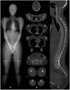
Figure 1
Figure 1
(A) CT scout-view showing the correct patient position in the CT scanner to avoid beam-hardening artifacts on thoracic and lumbar spine. (B) Whole-body low-dose CT examination performed from the skull down to the lower limbs. (C) Sagittal CT image showing cervical, dorsal and lumbar spine with “bone” algorithm is the best plane to identify vertebral compression fractures.
Figure 2
Figure 2
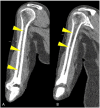
Coronal CT image showing (yellow arrowheads) humeral abnormal medullary lesions with high CT density in a patient with MM with (A) diffuse and (B) focal pattern.
Figure 3
Figure 3
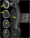
Multiplanar CT images showing (yellow arrowheads) osteolytic lesions of (A) the skull and of (B) a dorsal vertebra (detail in (B)).
Figure 4
Figure 4
Coronal CT image of (yellow arrowheads) bilateral femoral focal intra-medullary high CT density lesions in a patient with MM.
Figure 5
Figure 5
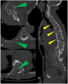
Multiplanar CT images showing humeral, ribs, and vertebral osteolytic lesions (green arrowheads in (AC)); multiple osteolytic lesions of the spine (yellow arrowheads in (D)).
Figure 6
Figure 6
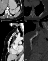
Multiplanar CT images showing (A) an osteolytic lesion of a rib, with (B) development of sclerosis and size-reduction after treatment. (C) Diffuse hyperdense myeloma deposits in the medullary cavities of the sternum with (D) development of sclerosis and size-reduction after treatment.
Figure 7
Figure 7
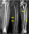
Coronal CT reconstruction images of the femoral bone showing: (A) regular aspect of bone marrow without osteolytic lesions, endosteal scalloping and pathological deposits; (B) “bone” algorithm reconstruction, used to evaluate osteolytic lesions and endosteal scalloping (yellow arrowheads); (C) “soft tissue” algorithm reconstruction used to assess focal and diffuse hyperdense deposits in the medullary cavities of long bones (yellow arrowheads).
Figure 8
Figure 8
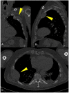
Multiplanar CT images showing a large para-medullary lesion (yellow arrowheads in (AC)): extra-osseous pathologic tissue in the chest wall soft tissues originating linked to skeletal involvement of the rib.
Figure 9
Figure 9
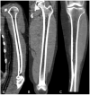
Multiplanar CT images with “soft tissue” window showing the distinctive aspect of normal (A) humeral, (B) femoral, and (C) tibial fatty bone marrow on low-dose CT in healthy adults.
Figure 10
Figure 10
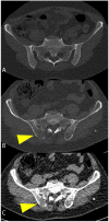
(A) Axial CT images showing normal appearance of bone and bone marrow in an axial skeleton with dense trabeculae. (B,C) myeloma-related osteolysis appearing as focal destructive lesions of the trabecular bone with cortical interruption and without a sclerotic border (yellow arrowheads).
Figure 11
Figure 11
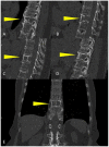
Sagittal CT reconstruction from right to left (AD), and (E) coronal multiplanar reconstruction of a dorsal vertebra showing a large osteolytic lesion involving more than 50% of the vertebral body (yellow arrowheads), with high risk of fracture.

References

    1. Hwang S., Panicek D.M. Magnetic resonance imaging of bone marrow in oncology, Part 1. Skelet. Radiol. 2007;36:913–920. doi: 10.1007/s00256-007-0309-3. - DOI - PubMed
    1. Małkiewicz A., Dziedzic M. Bone marrow reconversion—Imaging of physiological changes in bone marrow. Pol. J. Radiol. 2012;77:45–50. doi: 10.12659/PJR.883628. - DOI - PMC - PubMed
    1. Hillengass J., Usmani S., Rajkumar S.V., Durie B.G.M., Mateos M.V., Lonial S., Joao C., Anderson K.C., García-Sanz R., Riva E., et al. International myeloma working group consensus recommendations on imaging in monoclonal plasma cell disorders. Lancet Oncol. 2019;20:e302–e312. doi: 10.1016/S1470-2045(19)30309-2. - DOI - PubMed
    1. Rajkumar S.V., Dimopoulos M.A., Palumbo A., Blade J., Merlini G., Mateos M.V., Kumar S., Hillengass J., Kastritis E., Richardson P., et al. International Myeloma Working Group updated criteria for the diagnosis of multiple myeloma. Lancet Oncol. 2014;15:e538–e548. doi: 10.1016/S1470-2045(14)70442-5. - DOI - PubMed
    1. Fairfield H., Falank C., Avery L., Reagan M.R. Multiple myeloma in the marrow: Pathogenesis and treatments. Ann. N. Y. Acad. Sci. 2016;1364:32–51. doi: 10.1111/nyas.13038. - DOI - PMC - PubMed

LinkOut - more resources