. 2021 Dec 9;22(24):13265.
doi: 10.3390/ijms222413265.

Nano-Sized Extracellular Matrix Particles Lead to Therapeutic Improvement for Cutaneous Wound and Hindlimb Ischemia

Affiliations

Nano-Sized Extracellular Matrix Particles Lead to Therapeutic Improvement for Cutaneous Wound and Hindlimb Ischemia

Sang Su Ha et al. Int J Mol Sci. .

Abstract

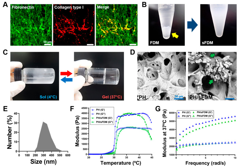
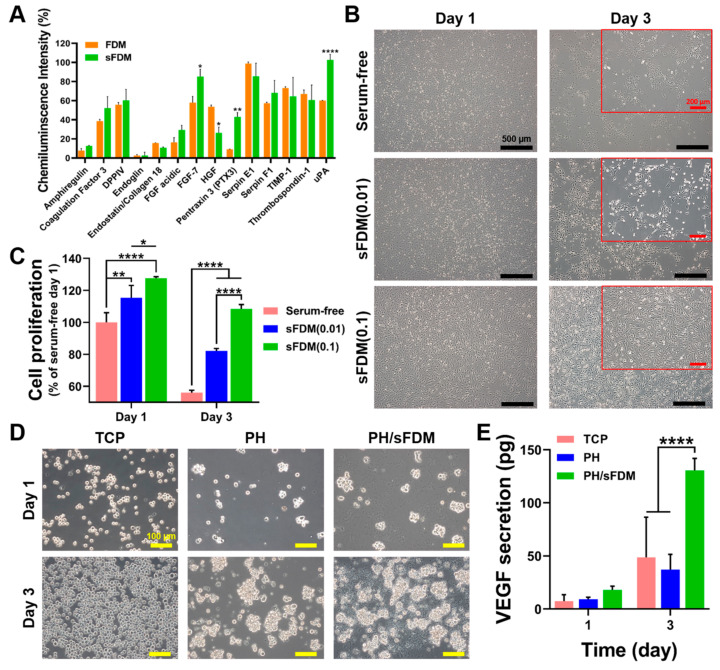
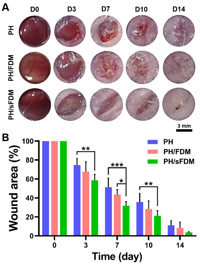
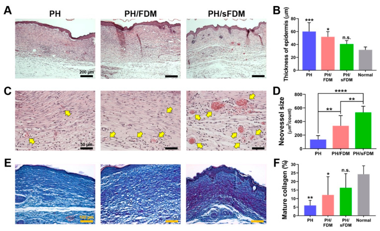
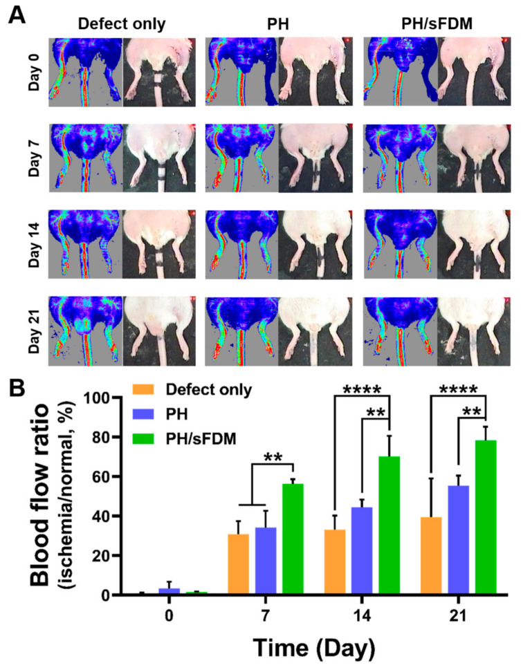
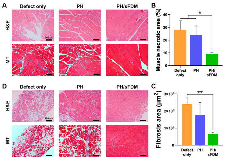
Cell-derived matrix (CDM) has proven its therapeutic potential and been utilized as a promising resource in tissue regeneration. In this study, we prepared a human fibroblast-derived matrix (FDM) by decellularization of in vitro cultured cells and transformed the FDM into a nano-sized suspended formulation (sFDM) using ultrasonication. The sFDM was then homogeneously mixed with Pluronic F127 and hyaluronic acid (HA), to effectively administer sFDM into target sites. Both sFDM and sFDM containing hydrogel (PH/sFDM) were characterized via immunofluorescence, sol-gel transition, rheological analysis, and biochemical factors array. We found that PH/sFDM hydrogel has biocompatible, mechanically stable, injectable properties and can be easily administered into the external and internal target regions. sFDM itself holds diverse bioactive molecules. Interestingly, sFDM-containing serum-free media helped maintain the metabolic activity of endothelial cells significantly better than those in serum-free condition. PH/sFDM also promoted vascular endothelial growth factor (VEGF) secretion from monocytes in vitro. Moreover, when we evaluated therapeutic effects of PH/sFDM via the murine full-thickness skin wound model, regenerative potential of PH/sFDM was supported by epidermal thickness, significantly more neovessel formation, and enhanced mature collagen deposition. The hindlimb ischemia model also found some therapeutic improvements, as assessed by accelerated blood reperfusion and substantially diminished necrosis and fibrosis in the gastrocnemius and tibialis muscles. Together, based on sFDM holding a strong therapeutic potential, our engineered hydrogel (PH/sFDM) should be a promising candidate in tissue engineering and regenerative medicine.

Keywords: Pluronic F127; decellularization; fibroblast-derived matrix (FDM); hindlimb ischemia; hyaluronic acid; suspended FDM; wound healing.


Conflict of interest statement

The authors declare no conflict of interest.

Figures

Figure 1
Figure 1
Preparation of suspended fibroblast-derived matrix (sFDM) and hydrogels and their characterizations. (A) ECM proteins in the decellularized FDM via immunofluorescence staining: fibronectin (green) and collagen type I (red). (B) Appearance of FDM before and after (sFDM) ultrasonication. Yellow arrow shows the collected FDM from the culture plate. (C) Sol–gel transition behavior of the hydrogel (PH) at 4 and 37 °C. (D) Internal microstructure of PH and PH/sFDM hydrogels via SEM. Green arrows indicate sFDM particles. (E) Measurement of particle size distribution of sFDM. (F) Profile of thermosensitive gelation between 20 and 45 °C for both hydrogels (PH and PH/sFDM) taken at 6.28 rad/s. (G) Comparison of both storage (G’) and loss (G”) moduli between the two hydrogels at body temperature.
Figure 2
Figure 2
Examination of biological properties of sFDM in vitro. (A) Quantitative assessment of angiogenic factors profile of both FDM and sFDM. (B) Human umbilical vein endothelial cells (HUVEC) morphology and cell adhesion when treated with three different types of media on day 1 and 3. Red boxes show the enlarged images of cell morphology at 3 days. (C) Quantitative analysis of cell proliferation after three different treatments for up to 3 days as compared to that of serum-free group. (D) Cultivation of THP-1 cells on the TCP, PH, and PH/sFDM. (E) Determination of VEGF released from each group at specific time points (day 1 and 3). Statistically significant difference (* p < 0.05, ** p < 0.01, **** p < 0.0001).
Figure 3
Figure 3
Observation of full-thickness wound closure with time. (A) Representative appearance of wounds treated with three test groups (PH, PH/FDM, PH/sFDM) on day 0, 3, 7, 10, and 14 post-surgery. (B) Quantitatively measured wound area ratio (%) at specific time points. Statistically significant difference (* p < 0.05, ** p < 0.01, *** p < 0.001).
Figure 4
Figure 4
Histological analysis of the wound tissues on day 14. (A) Representative images of the regenerated skin tissues via hematoxylin and eosin (H&E) staining. (B) Measurement of epidermal thickness as statistically compared to that of normal (* p < 0.05, *** p < 0.001), n.s. means no significance. (C) High resolution images in the dermis. Yellow arrows indicate neovessels. (D) Quantitative evaluation of newly formed vessel size. Statistically significant difference (** p < 0.01, **** p < 0.0001). (E) Herovici staining of the regenerated tissues. Blue and purple color in the dermis represent young and mature collagen fibers, respectively. (F) Quantitative analysis of mature collagen deposition. Statistically significant difference compared to that of normal (* p < 0.05, ** p < 0.01).
Figure 5
Figure 5
Blood perfusion recovery of ischemic hindlimb after the treatments for up to 21 days. (A) Representative images of laser Doppler perfusion imaging (LDPI) on day 0, 7, 14 and 21 post-treatments. Blue or dark-blue color indicates low and no perfusion, whereas red or yellow color indicates a high perfusion state. (B) Blood perfusion ratio was evaluated by comparing ischemic limb groups (defect only, PH, and PH/sFDM) with that of normal limb. Statistically significant difference (** p < 0.01, **** p < 0.0001).
Figure 6
Figure 6
Histological analysis of ischemic hindlimb muscles at 21 days post-treatments. (A) Necrosis and fibrosis in the gastrocnemius muscles was examined using H&E and Masson’s trichrome (MT) staining, respectively. (B) Quantitative analysis of the necrotic area and (C) the fibrosis area as suggested in purple color. (D) Fibrosis area in the tibialis muscles was examined using H&E and MT staining. Statistically significant difference (* p < 0.05, ** p < 0.01).

References

    1. Patan S. Vasculogenesis and angiogenesis as mechanisms of vascular network formation, growth and remodeling. J. Neuro-Oncol. 2000;50:1–15. doi: 10.1023/A:1006493130855. - DOI - PubMed
    1. Carmeliet P. Mechanisms of angiogenesis and arteriogenesis. Nat. Med. 2000;6:389–395. doi: 10.1038/74651. - DOI - PubMed
    1. Wu Y., Chen L., Scott P.G., Tredget E.E. Mesenchymal stem cells enhance wound healing through differentiation and angiogenesis. Stem Cells. 2007;25:2648–2659. doi: 10.1634/stemcells.2007-0226. - DOI - PubMed
    1. Deng C., Zhang P., Vulesevic B., Kuraitis D., Li F., Yang A.F., Griffith M., Ruel M., Suuronen E.J. A collagen–chitosan hydrogel for endothelial differentiation and angiogenesis. Tissue Eng. Part A. 2010;16:3099–3109. doi: 10.1089/ten.tea.2009.0504. - DOI - PubMed
    1. Jang W.B., Ji S.T., Park J.H., Kim Y.-J., Kang S., Lee N.-K., Kim J.S., Lim H.J., Choi J., Van Le T.H. Engineered M13 peptide carrier promotes angiogenic potential of patient-derived human cardiac progenitor cells and in vivo engraftment. Tissue Eng. Regen. Med. 2020;17:323–333. doi: 10.1007/s13770-020-00244-w. - DOI - PMC - PubMed

MeSH terms

LinkOut - more resources