Clinical Trial
. 2021 Dec 10;22(24):13294.
doi: 10.3390/ijms222413294.

The Study of a 231 French Patient Cohort Significantly Extends the Mutational Spectrum of the Two Major Usher Genes MYO7A and USH2A

Affiliations
Clinical Trial

The Study of a 231 French Patient Cohort Significantly Extends the Mutational Spectrum of the Two Major Usher Genes MYO7A and USH2A

Luke Mansard et al. Int J Mol Sci. .

Abstract

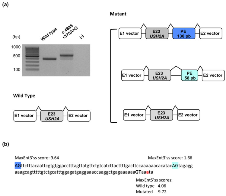
Usher syndrome is an autosomal recessive disorder characterized by congenital hearing loss combined with retinitis pigmentosa, and in some cases, vestibular areflexia. Three clinical subtypes are distinguished, and MYO7A and USH2A represent the two major causal genes involved in Usher type I, the most severe form, and type II, the most frequent form, respectively. Massively parallel sequencing was performed on a cohort of patients in the context of a molecular diagnosis to confirm clinical suspicion of Usher syndrome. We report here 231 pathogenic MYO7A and USH2A genotypes identified in 73 Usher type I and 158 Usher type II patients. Furthermore, we present the ACMG classification of the variants, which comprise all types. Among them, 68 have not been previously reported in the literature, including 12 missense and 16 splice variants. We also report a new deep intronic variant in USH2A. Despite the important number of molecular studies published on these two genes, we show that during the course of routine genetic diagnosis, undescribed variants continue to be identified at a high rate. This is particularly pertinent in the current era, where therapeutic strategies based on DNA or RNA technologies are being developed.

Keywords: ACMG classification; MYO7A; USH2A; Usher syndrome; deep intronic variant; hearing loss; pathogenic genotype; retinitis pigmentosa.


Conflict of interest statement

The authors declare no conflict of interest.

Figures

Figure A1
Figure A1
Genomic and encoded protein structure of MYO7A. (a) Genomic representation of the MYO7A gene and the associated Pfam (the protein families database; https://pfam.xfam.org) domains as indicated by UCSC Genome Browser (https://tinyurl.com/fbjwskcz, accessed on 8 December 2021). (b) Schematic representation of the UNIPROT myosin VIIa domains (Q13402, 2215 residues) and their localization within the protein.
Figure A2
Figure A2
Genomic and encoded protein structure of USH2A. (a) UCSC Genomic representation of the USH2A gene and the associated Pfam (the protein families database; https://pfam.xfam.org) domains as indicated by UCSC Genome Browser (https://tinyurl.com/2p9aexr5 accessed on 8 December 2021). (b) Schematic representation of the UNIPROT usherin domains (O75445, 5202 residues) and their distribution along the protein.
Figure 1
Figure 1
Types of variants in MYO7A (a) and USH2A (b) and proportion of novel alterations per type. The number of newly identified (purple bars) or already reported (pink bars) variants per alteration type are indicated within the bars. A total of 74 variants were identified for MYO7A (a) and 151 variants for USH2A (b). All variants were counted once, independently of their frequency in the cohort.
Figure 2
Figure 2
Mutational spectrum in MYO7A (a) and USH2A (b). Pie charts showing the allelic frequencies for each alteration type in the cohort; 146 alleles for MYO7A (a) and 316 alleles for USH2A (b) The variants were counted each time they occurred in the cohort.
Figure 3
Figure 3
Minigene assay of the c.4885+375A>G USH2A variant. (a) Electrophoretic visualization of RT-PCR products amplified from wild-type and mutant constructs and schematic representation of the corresponding splicing patterns. White boxes represent the pSPL3 exons, grey boxes correspond to exon 23 of USH2A and dark or light blue boxes represent two pseudoexons (PEs) of 130 and 58 nucleotides, respectively. (b) Intronic sequence involved in the inclusion of the two observed pseudoexons. The c.4885 adenine is indicated with red font; the activated donor splice site is shown with bold black font and the two used acceptor splice sites are highlighted in dark or light blue. MaxEnt donor splice site strength scores (5′ss scores) and MaxEnt acceptor splice site strength scores (3′ss scores) are indicated for the three splice sites.
Figure 4
Figure 4
Genotype spectrum of MYO7A (a) and USH2A (b). Pie charts on the left showing the frequencies of the different allele combinations. Pie charts on the right showing the number of patients carrying minor combinations of alleles.

References

    1. Millán J.M., Aller E., Jaijo T., Blanco-Kelly F., Gimenez-Pardo A., Ayuso C. An Update on the Genetics of Usher Syndrome. J. Ophthalmol. 2011;2011:1–8. doi: 10.1155/2011/417217. - DOI - PMC - PubMed
    1. Le Quesne Stabej P., Saihan Z., Rangesh N., Steele-Stallard H.B., Ambrose J., Coffey A., Emmerson J., Haralambous E., Hughes Y., Steel K.P., et al. Comprehensive sequence analysis of nine Usher syndrome genes in the UK National Collaborative Usher Study. J. Med. Genet. 2012;49:27–36. doi: 10.1136/jmedgenet-2011-100468. - DOI - PMC - PubMed
    1. Roux A.-F., Faugère V., Vaché C., Baux D., Besnard T., Léonard S., Blanchet C., Hamel C., Mondain M., Gilbert-Dussardier B., et al. Four-Year Follow-up of Diagnostic Service in USH1 Patients. Investig. Opthalmol. Vis. Sci. 2011;52:4063–4071. doi: 10.1167/iovs.10-6869. - DOI - PubMed
    1. Bonnet C., Riahi Z., Chantot-Bastaraud S., Smagghe L., Letexier M., Marcaillou C., Lefèvre G.M., Hardelin J.-P., El-Amraoui A., Singh-Estivalet A., et al. An innovative strategy for the molecular diagnosis of Usher syndrome identifies causal biallelic mutations in 93% of European patients. Eur. J. Hum. Genet. 2016;24:1730–1738. doi: 10.1038/ejhg.2016.99. - DOI - PMC - PubMed
    1. Abadie C., Blanchet C., Baux D., Larrieu L., Besnard T., Ravel P., Biboulet R., Hamel C., Malcolm S., Mondain M., et al. Audiological findings in 100 USH2 patients. Clin. Genet. 2011;82:433–438. doi: 10.1111/j.1399-0004.2011.01772.x. - DOI - PubMed

Publication types

Supplementary concepts

LinkOut - more resources