. 2021 Dec 11;22(24):13321.
doi: 10.3390/ijms222413321.

Zinc Exposure Promotes Commensal-to-Pathogen Transition in Pseudomonas aeruginosa Leading to Mucosal Inflammation and Illness in Mice

Affiliations

Zinc Exposure Promotes Commensal-to-Pathogen Transition in Pseudomonas aeruginosa Leading to Mucosal Inflammation and Illness in Mice

Tong Wu et al. Int J Mol Sci. .

Abstract

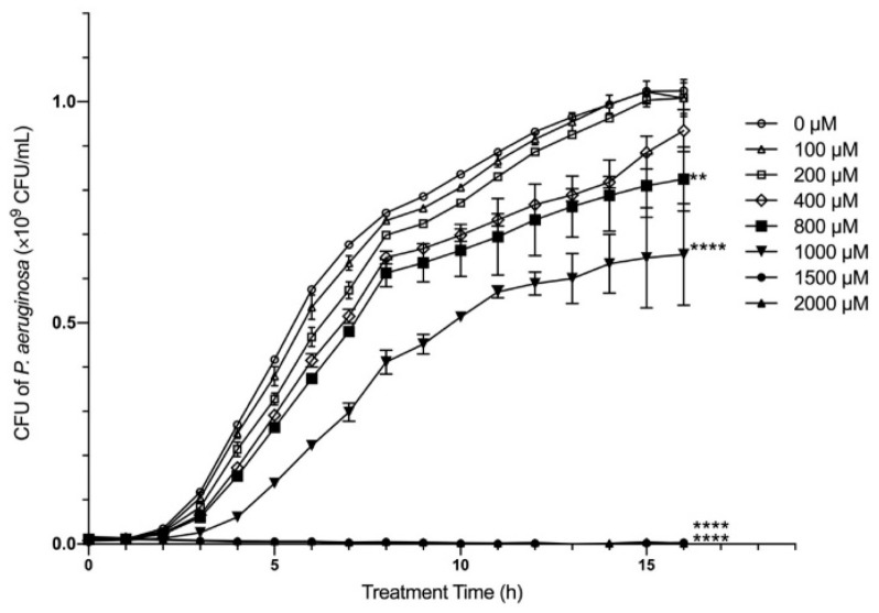
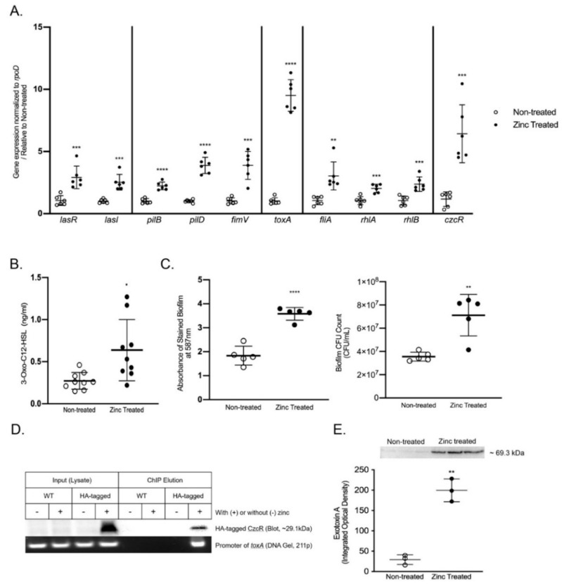
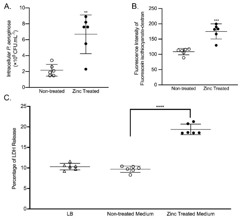
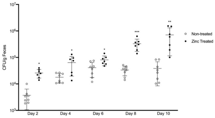
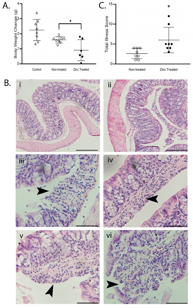
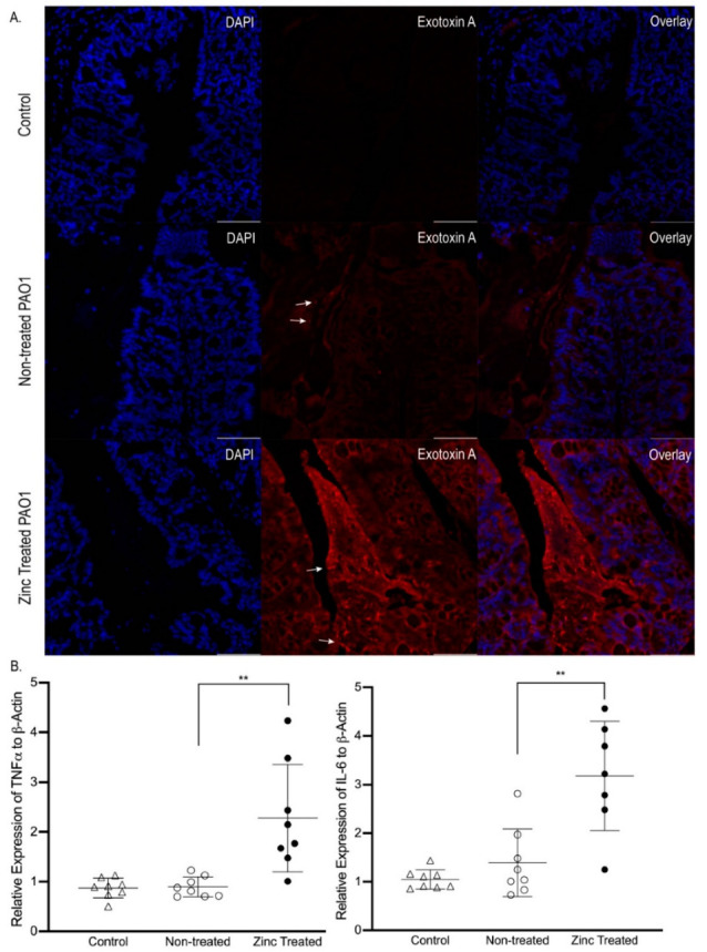
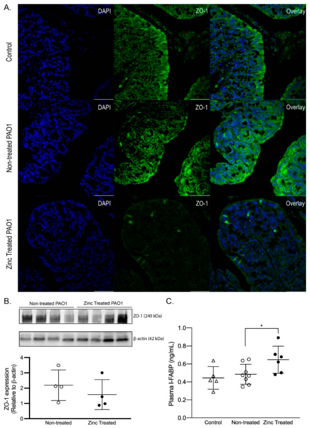
The opportunistic pathogen Pseudomonas aeruginosa (P. aeruginosa) is associated gastrointestinal (GI) inflammation and illness; however, factors motivating commensal-to-pathogen transition are unclear. Excessive zinc intake from supplements is common in humans. Due to the fact that zinc exposure enhances P. aeruginosa colonization in vitro, we hypothesized zinc exposure broadly activates virulence mechanisms, leading to inflammation and illness. P. aeruginosa was treated with excess zinc and growth, expression and secretion of key virulence factors, and biofilm production were determined. Effects on invasion, barrier function, and cytotoxicity were evaluated in Caco-2 cells co-cultured with P. aeruginosa pre-treated with zinc. Effects on colonization, mucosal pathology, inflammation, and illness were evaluated in mice infected with P. aeruginosa pre-treated with zinc. We found the expression and secretion of key virulence factors involved in quorum sensing (QS), motility (type IV pili, flagella), biosurfactants (rhamnolipids), toxins (exotoxin A), zinc homeostasis (CzcR), and biofilm production, were all significantly increased. Zinc exposure significantly increased P. aeruginosa invasion, permeability and cytotoxicity in Caco-2 cells, and enhanced colonization, inflammation, mucosal damage, and illness in mice. Excess zinc exposure has broad effects on key virulence mechanisms promoting commensal-to-pathogen transition of P. aeruginosa and illness in mice, suggesting excess zinc intake may have adverse effects on GI health in humans.

Keywords: Caco-2 cells; Pseudomonas aeruginosa; colonic inflammation; intestinal permeability; virulence; zinc.


Conflict of interest statement

All authors declare they have no competing interest.

Figures

Figure 1
Figure 1
The effect of zinc on P. aeruginosa growth. In vitro growth of PAO1 in the presence of ZnSO4 (0–2000 µM) was measured every hour for 16 h. The data shown are from one representative experiment, using three independent samples/groups. The data represent the mean CFU × 109/mL of PAO1 ± SD. ** p < 0.01, **** p < 0.0001 using Kruskal–Wallis. The experiment was repeated twice.
Figure 2
Figure 2
The effect of zinc on P. aeruginosa virulence. PAO1 was exposed to ZnSO4 (800 µM) for 16 h (zinc treated) and effects on key virulence pathways were compared to P. aeruginosa that was not exposed to excess zinc (non-treated). (A) Virulence gene expression. The data shown were generated from two independent experiments, using three independent samples/groups in each experiment. The data represent mean gene expression relative to rpoD ± SD, normalized to non-treated PAO1. *** p < 0.001, **** p < 0.0001 by student t test or ** p < 0.01 by Mann–Whitney. (B) Quantitation of 3-Oxo-C12 HSL in conditioned medium. The data shown were generated from three independent experiments, using three independent samples/groups in each experiment. The data represent the mean concentration of 3-Oxo-C12 HSL (ng/mL) ± SD. * p < 0.05 by student t test. (C) Biofilm formation. The data shown are from one representative experiment, using five independent samples/groups. The data represent the mean absorbance at 587 nm ± SD and the mean CFU/mL ± SD. ** p < 0.01, **** p < 0.0001 by student t test. The experiment was repeated twice. (D) Chromatin immunoprecipitation. Representative images of an immunoblot of hemagglutinin (HA; top) and an ethidium bromide-stained agarose gel (bottom). After verifying that HA-tagged CzcR protein was successfully expressed (Input Lysate; HA-tagged +) and precipitated by immunoblotting (ChIP elution; HA-tagged +), PCR was used to amplify the promoter region of toxA in CzcR-HA PAO1 treated for 16 h with zinc. Zinc-treated (WT; +) and non-treated PAO1 (WT; −), and non-zinc treated CzcR-HA PAO1 (HA-tagged; −) were used as negative controls. The ChIP experiment was repeated three times, using three independent samples. (E) Quantification of exotoxin A in the conditioned medium. Representative immunoblot of exotoxin A detected in the conditioned medium from PAO1 treated with (zinc treated) and without (non-treated) zinc. The data shown are from one representative experiment, using three independent samples/groups. The data represent the mean integrated optical density ± SD. ** p < 0.01 by student t test. The experiment was repeated twice.
Figure 3
Figure 3
The effect of zinc-exposed P. aeruginosa on Caco-2 cells. (A) Zinc-exposed P. aeruginosa increased invasive capacity. Caco-2 cells were co-cultured for 4 h with PAO1 previously exposed to ZnSO4 (800 µM) for 16 h (zinc treated) or non-treated PAO1, and extracellular bacteria were killed by gentamicin. Caco-2 cells were lysed, and the intracellular bacteria were enumerated by plating onto LB Agar. The data shown were generated from two independent experiments, using three independent samples/groups. The data represent the mean CFU × 105/mL ± SD. ** p < 0.01 by student t-test. (B) Zinc-exposed P. aeruginosa increased Caco-2 cell monolayer permeability. Caco-2 cells were co-cultured for 12 h with PAO1 previously exposed to ZnSO4 (800 µM) for 16 h (zinc treated) or non-treated PAO1. Fluorescein isothiocyanate-dextran was added to the apical chamber. The medium in the basolateral chamber was collected, and the fluorescence intensity was measured. The data shown were generated from two independent experiments, using three independent samples/groups. The data represent the mean fluorescence intensity ± SD. *** p < 0.001 by student t-test. (C) Zinc-exposed P. aeruginosa increased cell death. Caco-2 cells were co-cultured for 6 h with conditioned medium from PAO1 previously exposed to ZnSO4 (800 µM) for 16 h (Zinc treated medium) or non-treated PAO1 medium. Caco-2 cells co-cultured with fresh LB for 6 h were used as a negative control. Cytotoxicity was determined by LDH assay. The data shown were generated from two independent experiments, using three independent samples/groups. The data represent the mean percentage of LDH released relative to total LDH ± SD. **** p < 0.0001 by ANOVA.
Figure 4
Figure 4
Zinc treatment increased the ability of P. aeruginosa to colonize the mouse GI tract. PAO1 was exposed to ZnSO4 (800 µM) for 16 h (Zinc treated) or left untreated (non-treated) and orally gavaged in mice. Colony forming units (CFU) from the feces of mice inoculated with zinc treated or non-treated PAO1 were quantified every two days by plating the feces on P. aeruginosa-selective cetrimide agar plates. The data represent the mean CFU/g feces ± SD, n = 7–8 mice/group. * p < 0.05, ** p < 0.01, *** p < 0.001, relative to non-treated PAO1 mice at each timepoint, by student t-test.
Figure 5
Figure 5
Infection with zinc-exposed P. aeruginosa causes illness in mice. PAO1 was exposed to ZnSO4 (800 µM) for 16 h (zinc treated) or left untreated (non-treated) and orally gavaged in mice who were fed a control diet for 2 weeks prior to oral gavage. Mice not gavaged with PAO1 were used for comparison (control). (A) Zinc-exposed P. aeruginosa decreased body weight gain. The data represent the mean body weight change (g) ± SD, n = 6–8 mice/group. * p < 0.05, relative to non-treated PAO1 mice, by ANOVA. Note that infection with non-treated PAO1 did not significantly decrease body weight. (B) Zinc-exposed P. aeruginosa caused colonic pathology. Representative images of H&E-stained colon sections. Note that the histology in control mice (i) and mice infected with non-treated PAO1 (ii) was similar and had no pathological manifestations. Architectural distortion was found only in colons of mice infected with zinc treated PAO1 (iiivi). Arrowheads identify classical histopathology including effacement (iii), neutrophil infiltration (iv), and ulcer formation (v,vi). Magnification, 10× for control and non-treated PAO1 images (scale bar, 100 µm); 40× for zinc treated PAO1 images to highlight architectural distortion (scale bar, 50 µm). (C) Zinc-exposed P. aeruginosa increased total illness score. Illness was scored using the following parameters: daily body weight loss; general behavior; stool consistency; stool blood; and colon epithelial morphology and inflammatory cell infiltration documented using H&E staining [42]. The data represent the mean total illness score ± SD, n = 8 mice/group. * p < 0.05 by student t-test.
Figure 6
Figure 6
Infection with zinc-exposed P. aeruginosa induced greater mucosal inflammatory responses in mice. PAO1 was exposed to ZnSO4 (800 µM) for 16 h (zinc treated) or left untreated (non-treated) and orally gavaged in mice. Mice not gavaged with PAO1 were used for comparison (control). (A) Mice infected with zinc-exposed P. aeruginosa had greater exotoxin A in the colon. Representative images of exotoxin A (red) and nuclei stained with DAPI (blue) in colon of control mice, and mice infected with Non-treated PAO1 or Zinc-treated PAO1. No exotoxin A was observed in control mice (negative control). In mice infected with Non-treated PAO1, little fluorescence was detected, and what was detected was observed as punctate rods, indicative of intact PAO1 (white arrows). In contrast, intense fluorescence was detected as both an overall haze throughout the tissue and as punctate rods (white arrows) in mice infected with Zinc treated PAO1. Additionally, the formation of biofilm structures was only detected in the colons of mice infected with Zinc treated PAO1 (note defined triangular structure between colonic folds). Magnification, 40×; Scale bar, 50 µm. (B) Zinc-exposed P. aeruginosa activated colon inflammation. Data represent mean fold change of TNFα, and IL-6 mRNA levels normalized to β-actin ± SD, relative to control mice, n = 7–8 mice/group. ** p < 0.01 by ANOVA. Note that infection with Non-treated PAO1 caused no increase in TNFα and IL-6 mRNA levels.
Figure 7
Figure 7
Zinc-exposed P. aeruginosa increased colon permeability. PAO1 was exposed to ZnSO4 (800 µM) for 16 h (Zinc treated) or left untreated (Non-treated) and orally gavaged in mice. Mice not gavaged with PAO1 were used for comparison (control). (A) Mice infected with zinc-exposed P. aeruginosa had disorganized ZO-1 localization in the colon. Representative images of ZO-1 (green) and nuclei stained with DAPI (blue) in the colons of control mice or mice infected with non-treated or zinc treated PAO1. Magnification, 40×; scale bar, 50 µm. (B) Zinc-exposed P. aeruginosa did not affect ZO-1 abundance in the colon. Representative immunoblots of ZO-1 in the colons of mice infected with non-treated or zinc treated PAO1. Membranes were stripped and re-probed for β-actin as a loading control. The data shown are from one representative experiment. The data represent the mean integrated optical density normalized to β-actin ± SD, n = 4 mice/group. The experiment was repeated twice, using four different mice/groups. (C) Zinc-exposed P. aeruginosa increased plasma I-FABP concentration. The data represent the mean concentration of I-FABP (ng/mL) ± SD, n = 5–8 mice/group. * p < 0.05, relative to non-treated PAO1 group, by ANOVA. Note that infection with non-treated PAO1 caused no increase in plasma I-FABP concentration.

References

    1. Yoshioka H., Fujita K., Maruyama S. Faecal carriage of Pseudomonas aeruginosa in newborn infants. J. Hosp. Infect. 1983;4:41–44. doi: 10.1016/0195-6701(83)90063-4. - DOI - PubMed
    1. Wagner J., Short K., Catto-Smith A.G., Cameron D.J., Bishop R.F., Kirkwood C.D. Identification and characterisation of Pseudomonas 16S ribosomal DNA from ileal biopsies of children with Crohn’s disease. PLoS ONE. 2008;3:e3578. doi: 10.1371/journal.pone.0003578. - DOI - PMC - PubMed
    1. Swidsinski A., Ladhoff A., Pernthaler A., Swidsinski S., Loening-Baucke V., Ortner M., Weber J., Hoffmann U., Schreiber S., Dietel M., et al. Mucosal flora in inflammatory bowel disease. Gastroenterology. 2002;122:44–54. doi: 10.1053/gast.2002.30294. - DOI - PubMed
    1. Kerckhoffs A.P., Ben-Amor K., Samsom M., van der Rest M.E., de Vogel J., Knol J., Akkermans L.M. Molecular analysis of faecal and duodenal samples reveals significantly higher prevalence and numbers of Pseudomonas aeruginosa in irritable bowel syndrome. J. Med. Microbiol. 2011;60:236–245. doi: 10.1099/jmm.0.022848-0. - DOI - PubMed
    1. Chuang C.-H., Janapatla R.P., Wang Y.-H., Chang H.-J., Huang Y.-C., Lin T.-Y., Chiu C.-H. Pseudomonas aeruginosa-associated diarrheal diseases in children. Pediatric Infect. Dis. J. 2017;36:1119–1123. doi: 10.1097/INF.0000000000001567. - DOI - PubMed

LinkOut - more resources