. 2021 Dec 14;22(24):13444.
doi: 10.3390/ijms222413444.

A Complex Evaluation of the In-Vivo Biocompatibility and Degradation of an Extruded ZnMgSr Absorbable Alloy Implanted into Rabbit Bones for 360 Days

Affiliations

A Complex Evaluation of the In-Vivo Biocompatibility and Degradation of an Extruded ZnMgSr Absorbable Alloy Implanted into Rabbit Bones for 360 Days

Karel Klíma et al. Int J Mol Sci. .

Abstract

The increasing incidence of trauma in medicine brings with it new demands on the materials used for the surgical treatment of bone fractures. Titanium, its alloys, and steel are used worldwide in the treatment of skeletal injuries. These metallic materials, although inert, are often removed after the injured bone has healed. The second-stage procedure-the removal of the plates and screws-can overwhelm patients and overload healthcare systems. The development of suitable absorbable metallic materials would help us to overcome these issues. In this experimental study, we analyzed an extruded Zn-0.8Mg-0.2Sr (wt.%) alloy on a rabbit model. From this alloy we developed screws which were implanted into the rabbit tibia. After 120, 240, and 360 days, we tested the toxicity at the site of implantation and also within the vital organs: the liver, kidneys, and brain. The results were compared with a control group, implanted with a Ti-based screw and sacrificed after 360 days. The samples were analyzed using X-ray, micro-CT, and a scanning electron microscope. Chemical analysis revealed only small concentrations of zinc, strontium, and magnesium in the liver, kidneys, and brain. Histologically, the alloy was verified to possess very good biocompatibility after 360 days, without any signs of toxicity at the site of implantation. We did not observe raised levels of Sr, Zn, or Mg in any of the vital organs when compared with the Ti group at 360 days. The material was found to slowly degrade in vivo, forming solid corrosion products on its surface.

Keywords: absorbable metals; alloy accumulation; biocompatibility; in vivo; internal organs; magnesium; strontium; systemic reactions; toxicity; zinc.


Conflict of interest statement

The authors declare no conflict of interest.

Figures

Figure 1
Figure 1
X-ray images (plain radiography) of the tibia bones of experimental rabbits. (ad) 120 days after implantation, (ac) experimental Zn-based screw, (d) control Ti-based screw; (eg) 240 days after implantation; (e,f) experimental Zn-based screw; (g) control Ti-based screw; (h) 360 days after experimental Zn-based screw implantation; (i) 360 days after control Ti-based screw implantation. There were no osteolytic changes seen on the boundaries between the bone and experimental and Ti-based screws. No detectable differences between groups after 120, 240, and 360 days of implantation. No detectable changes between experimental screw groups and Ti-based screws used as a control.
Figure 2
Figure 2
Micro-CT visualizations; (a) standardized cross-sectional images in 3 perpendicular planes; (b) implant surrounded by bone; (c) 3D visualization of implant 240 days after healing. Scale bar = 4 mm.
Figure 3
Figure 3
Micro-CT cross-sectional images; (a) tested implant, 120 days of healing; (b) tested implant, 240 days of healing; (c) tested implant, 360 days of healing; (d) control specimen (Ti-based screw). All specimens showed bone healing without any signs of adverse reaction. To enhance clarity, image noise was reduced except around the areas of the bone and implant. Scale bar = 4 mm.
Figure 4
Figure 4
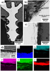
(ad) SEM micrographs (Z-contrast) of a Zn-0.8Mg-0.2Sr experimental screw 240 days after implantation in rabbit tibias. The X-ray elemental maps showing the distributions of Zn, Ca, P, O, and C were acquired from the area shown in (d); the scale bar is the same as in (d). In (b) and (c), the gray shaded areas can be interpreted as follows: the lightest gray—metallic implant, light gray—corrosion products, dark gray—bone tissue and black/the darkest gray—fibrous tissue.
Figure 5
Figure 5
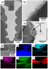
(ad) SEM micrographs (Z-contrast) of a Zn-0.8Mg-0.2Sr experimental screw 360 days after implantation in rabbit tibias. The X-ray elemental maps showing the distributions of Zn, Ca, P, O, and C were acquired from the area shown in (d); the scale bar is the same as in (d). In (b,c), the gray shaded areas can be interpreted as follows: the lightest gray—metallic implant, light gray—corrosion products, dark gray—bone tissue and black/the darkest gray—fibrous tissue.
Figure 6
Figure 6
(ad) SEM micrographs (Z-contrast) of a Ti-based control screw 360 days after implantation in rabbit tibias. The X-ray elemental maps showing the distributions of Ti, Al, V, Ca, P, O, and C were acquired from the area shown in (d); the scale bar is the same as in (d). In (b,c), the gray shaded areas can be interpreted as follows: the lightest gray—metallic implant, dark gray—bone tissue and black/the darkest gray—fibrous tissue.
Figure 7
Figure 7
Histological examination of bone specimens (af) 120 days after experimental screw implantation; (g,h) 240 days after experimental Zn-based screw implantation; (i,j) 360 days after experimental screw implantation. Mild changes in the experimental screw sharpness, suggesting possible corrosion (k,l) 360 days after Ti-based screw implantation. Osteon-like cavities—arrows. Figure 7a,c,d are assumed from our previous pilot study [18].
Figure 8
Figure 8
Peri-implant bone formation (arrows), variable thickness of newly formed bone on the surface of the implanted screws (marked by the yellow arrows) (a) 120 days after experimental screw implantation, (b) 240 days after experimental screw implantation, (c) 360 days after experimental screw implantation. (d) Control 360 days after Ti-based screw implantation.
Figure 9
Figure 9
Peri-implant fibrosis (fibrosis is marked by yellow arrows) (a) 120 days after experimental screw implantation, (b) 240 days after experimental screw implantation, (c) 360 days after experimental screw implantation, (d) 360 days after Ti-based screw implantation.
Figure 10
Figure 10
Peri-implant inflammation (marked by yellow arrows) (a) 120 days after experimental screw implantation, (b) 240 days after experimental screw implantation, (c) 360 days after experimental screw implantation, (d) 360 days after Ti-based screw implantation.
Figure 11
Figure 11
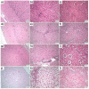
Histological analysis of vital organs, brain (left column), liver (middle column), and kidney (right column) (ac) 120 days after experimental screw implantation, (df), 240 days after experimental screw implantation, (gi) 360 days after experimental screw implantation, (jl) 360 days after Ti-based screw implantation. No detectable changes between experimental screw groups and Ti-based screws used as a control. (k) Massive hepatic steatosis visible in the liver occurred 360 days after Ti-based screw implantation.

References

    1. Burge R., Dawson-Hughes B., Solomon D.H., Wong J.B., King A., Tosteson A. Incidence and Economic Burden of Osteoporosis-Related Fractures in the United States, 2005–2025. J. Bone Miner. Res. 2007;22:465–475. doi: 10.1359/jbmr.061113. - DOI - PubMed
    1. Han H.-S., Loffredo S., Jun I., Edwards J., Kim Y.-C., Seok H.-K., Witte F., Mantovani D., Glyn-Jones S. Current Status and Outlook on the Clinical Translation of Biodegradable Metals. Mater. Today. 2019;23:57–71. doi: 10.1016/j.mattod.2018.05.018. - DOI
    1. Wang X., Shao X., Dai T., Xu F., Zhou J.G., Qu G., Tian L., Liu B., Liu Y. In Vivo Study of the Efficacy, Biosafety, and Degradation of a Zinc Alloy Osteosynthesis System. Acta Biomater. 2019;92:351–361. doi: 10.1016/j.actbio.2019.05.001. - DOI - PubMed
    1. Tan L., Yu X., Wan P., Yang K. Biodegradable Materials for Bone Repairs: A Review. J. Mater. Sci. Technol. 2013;29:503–513. doi: 10.1016/j.jmst.2013.03.002. - DOI
    1. Gombotz W.R., Pettit D.K. Biodegradable Polymers for Protein and Peptide Drug Delivery. Bioconjugate Chem. 1995;6:332–351. doi: 10.1021/bc00034a002. - DOI - PubMed

MeSH terms