

A Comparative Study of Koizumi and Longa Methods of Intraluminal Filament Middle Cerebral Artery Occlusion in Rats: Early Corticosterone and Inflammatory Response in the Hippocampus and Frontal Cortex
- PMID: 34948340
- PMCID: PMC8703333
- DOI: 10.3390/ijms222413544
A Comparative Study of Koizumi and Longa Methods of Intraluminal Filament Middle Cerebral Artery Occlusion in Rats: Early Corticosterone and Inflammatory Response in the Hippocampus and Frontal Cortex
Abstract
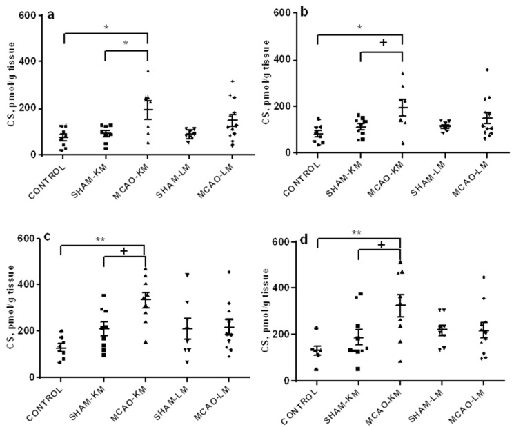
Two classical surgical approaches for intraluminal filament middle cerebral artery occlusion (MCAO), the Longa et al. (LM) and Koizumi et al. methods (KM), are used as alternatives in preclinical studies to induce stroke in rodents. Comparisons of these MCAO models in mice showed critical differences between them along with similarities (Smith et al. 2015; Morris et al. 2016). In this study, a direct comparison of MCAO-KM and MCAO-LM in rats was performed. Three days after MCAO, infarct volume, mortality rate, neurological deficit, and weight loss were similar in these models. MCAO-LM rats showed an increase in ACTH levels, while MCAO-KM rats demonstrated elevated corticosterone and interleukin-1β in blood serum. Corticosterone accumulation was detected in the frontal cortex (FC) and the hippocampus of the MCAO-KM group. IL1β beta increased in the ipsilateral hippocampus in the MCAO-KM group and decreased in the contralateral FC of MCAO-LM rats. Differences revealed between MCAO-KM and MCAO-LM suggest that corticosterone and interleukin-1β release as well as hippocampal accumulation is more expressed in MCAO-KM rats, predisposing them to corticosterone-dependent distant neuroinflammatory hippocampal damage. The differences between two models, particularly, malfunction of the hypothalamic-pituitary-adrenal axis, should be considered in the interpretation, comparison, and translation of pre-clinical experimental results.
Keywords: adrenocorticotropic hormone; corticosterone; distant damage; frontal cortex; hippocampus; interleukins; middle cerebral artery occlusion; neuroinflammation; stroke.
Conflict of interest statement
The authors declare no conflict of interest. The funders had no role in the design of the study; in the collection, analyses, or interpretation of data; in the writing of the manuscript, or in the decision to publish the results.
Figures


References
-
- Feigin V.L., Nichols E., Alam T., Bannick M.S., Beghi E., Blake N., Culpepper W.J., Dorsey E.R., Elbaz A., Ellenbogen R.G., et al. Global, regional, and national burden of neurological disorders, 1990–2016: A systematic analysis for the Global Burden of Disease Study. Lancet Neurol. 2019;18:459–480. doi: 10.1016/S1474-4422(18)30499-X. - DOI - PMC - PubMed
Publication types
MeSH terms
Substances
Grants and funding
LinkOut - more resources
Full Text Sources
Medical
