. 2021 Dec 20;22(24):13671.
doi: 10.3390/ijms222413671.

Luteolin Improves Perivascular Adipose Tissue Profile and Vascular Dysfunction in Goto-Kakizaki Rats

Affiliations

Luteolin Improves Perivascular Adipose Tissue Profile and Vascular Dysfunction in Goto-Kakizaki Rats

Marcelo Queiroz et al. Int J Mol Sci. .

Abstract

We investigated the effects of luteolin on metabolism, vascular reactivity, and perivascular adipose tissue (PVAT) in nonobese type 2 diabetes mellitus animal model, Goto-Kakizaki (GK) rats.

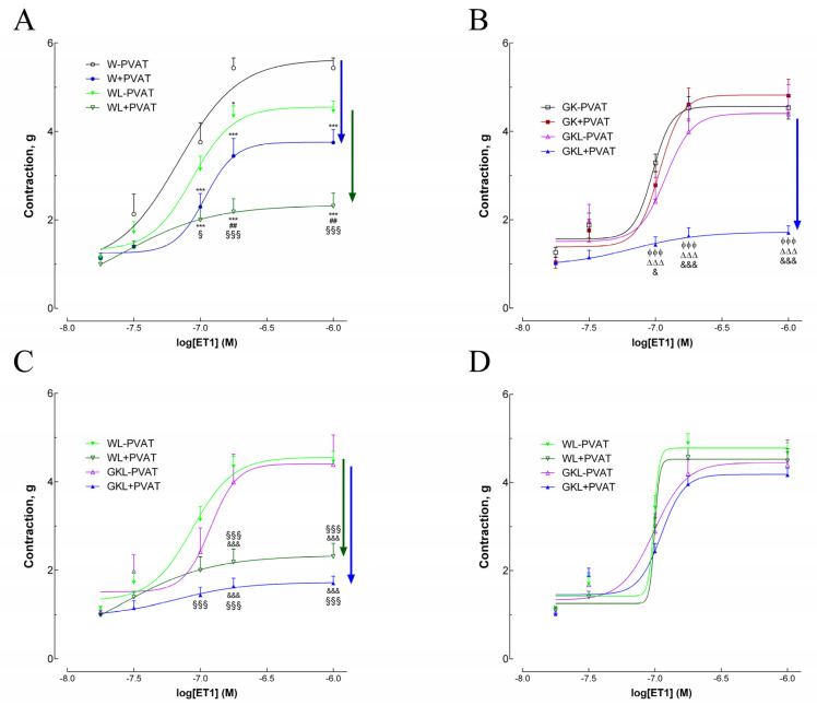
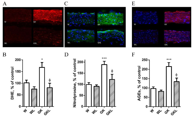
Methods: Wistar and GK rats were divided in two groups: (1) control groups treated with vehicle; (2) groups treated with luteolin (10 mg/kg/day, for 2 months). Several metabolic parameters such as adiposity index, lipid profile, fasting glucose levels, glucose and insulin tolerance tests were determined. Endothelial function and contraction studies were performed in aortas with (PVAT+) or without (PVAT-) periaortic adipose tissue. We also studied vascular oxidative stress, glycation and assessed CRP, CCL2, and nitrotyrosine levels in PVAT.

Results: Endothelial function was impaired in diabetic GK rats (47% (GK - PVAT) and 65% (GK + PVAT) inhibition of maximal endothelial dependent relaxation) and significantly improved by luteolin treatment (29% (GK - PVAT) and 22% (GK + PVAT) inhibition of maximal endothelial dependent relaxation, p < 0.01). Vascular oxidative stress and advanced glycation end-products' levels were increased in aortic rings (~2-fold, p < 0.05) of diabetic rats and significantly improved by luteolin treatment (to levels not significantly different from controls). Periaortic adipose tissue anti-contractile action was significantly rescued with luteolin administration (p < 0.001). In addition, luteolin treatment significantly recovered proinflammatory and pro-oxidant PVAT phenotype, and improved systemic and metabolic parameters in GK rats.

Conclusions: Luteolin ameliorates endothelial dysfunction in type 2 diabetes and exhibits therapeutic potential for the treatment of vascular complications associated with type 2 diabetes.

Keywords: endothelial dysfunction; inflammation; luteolin; oxidative stress; type 2 diabetes.


Conflict of interest statement

The authors declare no conflict of interest.

Figures

Figure 1
Figure 1
Influence of luteolin on blood glucose levels throughout an intraperitoneal glucose tolerance test (IPGTT; (A)), the glucose area under the curve (AUC; (B)), fasting glycemia (C), insulin tolerance test (ITT; D), the insulin area under the curve (AUC; (E)), and homeostasis model assessment of insulin resistance (HOMA, (F)) in normal Wistar (W), diabetic Goto-Kakizaki (GK) control rats and rats treated with luteolin (10 mg/kg/day, orally) for 2 months (WL, GKL). Data are expressed as mean ± SE (n = 10). * p < 0.05, ** p < 0.01, *** p < 0.001 vs. W group; ϕ p < 0.05, ϕϕ p < 0.01, ϕϕϕ p < 0.001 vs. GK group.
Figure 2
Figure 2
Effects of luteolin on vascular responses to acetylcholine (ACh, panel AC), and sodium nitroprusside (SNP, panel DF) in aortas of normal Wistar (W) and diabetic Goto-Kakizaki (GK) rats mounted with or without perivascular adipose tissue (PVAT). (A,B) Endothelium-dependent vasodilation in aortic rings in the absence (−PVAT) or presence of PVAT (+PVAT) was assessed in W (panel A) and GK rats (panel B) with or without luteolin treatment. (C) Comparison of endothelium-dependent vasodilation in aortic rings in W and GK rats treated with luteolin. (D). Vasodilator response to SNP in aortas of W (panel D) and GK (panel E) rats. Endothelium-independent vasodilation in aortic rings in the absence (−PVAT) or presence of PVAT (+PVAT) was assessed in W and GK with or without luteolin treatment (panel DF). (F) Comparison of endothelium-independent vasodilation in aortic rings in W and GK rats treated with luteolin. Data are expressed as mean ± SE (n = 10). * p < 0.05, ** p < 0.01, *** p < 0.001 vs. W-PVAT group; # p < 0.05, ## p < 0.01, ### p < 0.001 vs. W + PVAT group; § p < 0.05, §§ p < 0.01, §§§ p < 0.001 vs. WL-PVAT group; $ p < 0.05 vs. WL + PVAT group; ϕ p < 0.05, ϕϕ p < 0.01, ϕϕϕ p < 0.001 vs. GK-PVAT group;  p < 0.05, ∆∆ p < 0.01, ∆∆∆ p < 0.001 vs. GK + PVAT group; & p < 0.05, && p < 0.01, &&& p < 0.001 vs. GKL-PVAT.
Figure 3
Figure 3
Effects of luteolin on contraction responses to endothelin-1 (ET1) in aortas of Wistar (W) and diabetic Goto-Kakizaki (GK) rats in the absence (−PVAT) or presence of PVAT (+PVAT). The effect of luteolin is highlighted in panel (A) and (B) for W and GK aortas, respectively. Luteolin recovered the anticontractile PVAT phenotype typical of normal W in diabetic GK rats (panel B,C). (D) Effects of luteolin on contraction responses to ET1 in aortas of W and GK rats in the presence of 4-aminopiridine, a voltage-dependent potassium channel (Kv) inhibitor. Luteolin treatment promoted a substantial PVAT anticontractile effect in aortic rings (panel B,C) lost after treatment with 4-aminopiridine (panel D). Data are expressed as mean ± SE (n = 10). * p < 0.05, *** p < 0.001 vs. W-PVAT group; ## p < 0.01 vs. W + PVAT group; § p < 0.05, §§§ p < 0.001 vs. WL-PVAT group; ϕϕϕ p < 0.001 vs. GK-PVAT group; ∆∆∆ p < 0.001 vs. GK + PVAT group; & p < 0.05, &&& p < 0.001 vs. GKL-PVAT.
Figure 4
Figure 4
Effects of luteolin treatment on vascular oxidative stress and advanced glycation end- products’ levels in Wistar (W) and diabetic Goto-Kakizaki (GK) rats. Representative DHE-stained aorta artery sections reflect O2•− production in W and GK rats with (WL, GKL) or without luteolin treatment (W, GK). (Panel A). The endothelium is facing up in all layers. At identical settings, fluorescence (reflecting O2•− levels in the endothelium, intima, and media) in diabetic GK was markedly increased compared to age-matched control W rats. DHE fluorescence decreased in the diabetic GK rats treated with luteolin (GKL). Panel (B) contains quantification of the fluorescence ethidium signal in the different groups of arteries. Representative aortic sections showing nitrotyrosine staining (panel C) in W and GK rats with (WL, GKL) or without luteolin treatment (W, GK). Panel (D) contains quantification of the green fluorescence in the different groups of arteries. Representative aortic sections showing AGE staining (panel E) in W and GK rats with (WL, GKL) or without luteolin treatment (W, GK). Panel (F) exhibits quantification of the red fluorescence in the various groups of arteries. Data are expressed as mean ± SE (n = 10). * p < 0.05, *** p < 0.001 vs. W group; ϕ p < 0.05 vs. GK group.
Figure 5
Figure 5
Effects of luteolin treatment on reduced antioxidant defenses and increased lipid peroxidation in perivascular adipose tissue (PVAT) of thoracic aorta from diabetic Goto-Kakizaki (GK) rats. Activities of manganese superoxide dismutase (MnSOD; A) and aldose reductase (AR; B), tissue contents of glutathione (GSH; C), and malonaldehyde levels (MDA; D) in PVAT of thoracic aortas of GK rats compared with nondiabetic Wistar (W) rats treated with (WL, GKL) or without luteolin. Data are expressed as mean ± SE (n = 10). *** p < 0.001 vs. W group; ϕϕϕ p < 0.001 vs. GK group; ** p < 0.01 vs. W group; ϕϕ p < 0.01, vs. GK group.
Figure 6
Figure 6
Effects of luteolin on inflammation of perivascular adipose tissue (PVAT) in thoracic aorta from Wistar and Goto-Kakizaki (GK) rats. CRP (A) and CCL2 (B) levels; immunohistochemical staining for nitrotyrosine (C,D) were determined in PVAT of thoracic aortas of the different groups of rats. Data are expressed as mean ± SE. * p < 0.05, ** p < 0.01 vs. W group; ϕ p < 0.05 vs. GK group.

References

    1. Stehouwer C.D., Henry R.M., Ferreira I. Arterial stiffness in diabetes and the metabolic syndrome: A pathway to cardio-vascular disease. Diabetologia. 2008;51:527–539. doi: 10.1007/s00125-007-0918-3. - DOI - PubMed
    1. Okada S., Hiuge A., Makino H., Nagumo A., Takaki H., Konishi H., Goto Y., Yoshimasa Y., Miyamoto Y. Effect of exercise intervention on endothelial function and incidence of cardiovascular disease in patients with type 2 diabetes. J. Atheroscler. Thromb. 2010;17:828–833. doi: 10.5551/jat.3798. - DOI - PubMed
    1. Yahagi K., Kolodgie F.D., Lutter C., Mori H., Romero M.E., Finn A.V., Virmani R. Pathology of Human Coronary and Carotid Artery Atherosclerosis and Vascular Calcification in Diabetes Mellitus. Arter. Thromb. Vasc. Biol. 2017;37:191–204. doi: 10.1161/ATVBAHA.116.306256. - DOI - PMC - PubMed
    1. Sena C.M., Pereira A.M., Seiça R. Endothelial dysfunction—A major mediator of diabetic vascular disease. Biochim. Biophys. Acta (BBA)-Mol. Basis Dis. 2013;1832:2216–2231. doi: 10.1016/j.bbadis.2013.08.006. - DOI - PubMed
    1. Sena C.M., Matafome P., Louro T., Nunes E., Fernandes R., Seiça R.M. Metformin restores endothelial function in aorta of diabetic rats. Br. J. Pharmacol. 2011;163:424–437. doi: 10.1111/j.1476-5381.2011.01230.x. - DOI - PMC - PubMed

MeSH terms