Review
. 2022 Jan 4;119(1):e2111046118.
doi: 10.1073/pnas.2111046118.

Microenvironment-mediated cancer dormancy: Insights from metastability theory

Affiliations
Review

Microenvironment-mediated cancer dormancy: Insights from metastability theory

Sadra Bakhshandeh et al. Proc Natl Acad Sci U S A. .

Abstract

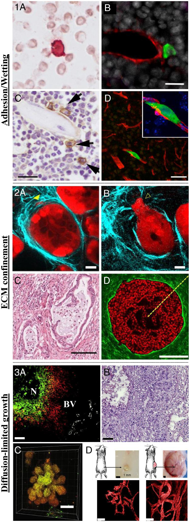
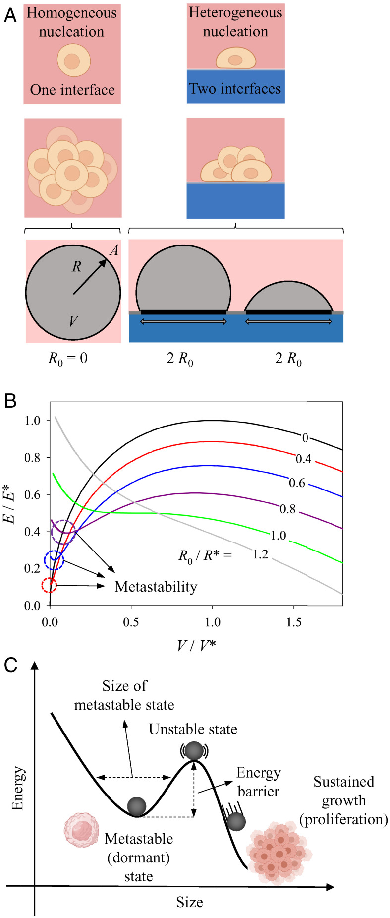
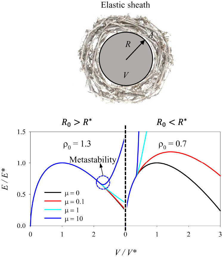
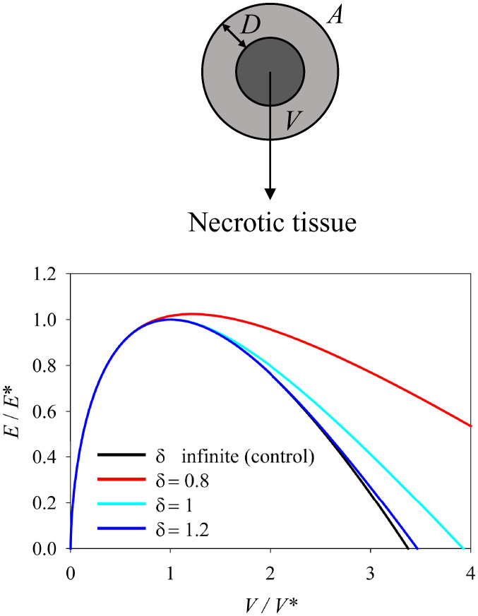
Dormancy is an evolutionarily conserved protective mechanism widely observed in nature. A pathological example is found during cancer metastasis, where cancer cells disseminate from the primary tumor, home to secondary organs, and enter a growth-arrested state, which could last for decades. Recent studies have pointed toward the microenvironment being heavily involved in inducing, preserving, or ceasing this dormant state, with a strong focus on identifying specific molecular mechanisms and signaling pathways. Increasing evidence now suggests the existence of an interplay between intracellular as well as extracellular biochemical and mechanical cues in guiding such processes. Despite the inherent complexities associated with dormancy, proliferation, and growth of cancer cells and tumor tissues, viewing these phenomena from a physical perspective allows for a more global description, independent from many details of the systems. Building on the analogies between tissues and fluids and thermodynamic phase separation concepts, we classify a number of proposed mechanisms in terms of a thermodynamic metastability of the tumor with respect to growth. This can be governed by interaction with the microenvironment in the form of adherence (wetting) to a substrate or by mechanical confinement of the surrounding extracellular matrix. By drawing parallels with clinical and experimental data, we advance the notion that the local energy minima, or metastable states, emerging in the tissue droplet growth kinetics can be associated with a dormant state. Despite its simplicity, the provided framework captures several aspects associated with cancer dormancy and tumor growth.

Keywords: cancer dormancy; extracellular matrix; metastability; phase separation; tissue growth.


Conflict of interest statement

The authors declare no competing interest.

Figures

Fig. 1.
Fig. 1.
Microenvironment-mediated cancer dormancy. (1) Adhesion/wetting. (1A) Disseminated breast cancer cell (red) in the proximity of neighboring cells in the bone marrow of patients with ductal carcinoma in situ (DCIS), the earliest and most noninvasive form of breast cancer (400-fold magnification). Adapted from ref. . (1B) Disseminated breast cancer cell (green) adhering to the microvasculature (red) of the bone marrow in a mouse model. (Scale bar, 20 µm.) Adapted from ref. . (1C) Disseminated MM cells (brown) adhere to the endosteal surface of trabecular bone in a mouse model. (Scale bar, 20 µm.) Adapted from ref. . (1D) Disseminated breast cancer cell (green) adhere to the microvasculature (red) of the brain in a mouse model. (Scale bar, 50 µm.) Adapted from ref. . (2) Mechanical confinement. (2A and 2B) Pancreatic ductal carcinoma (red) surrounded by collagen ECM (cyan) before (2A) and after (2B) invasion of the surrounding murine tissue. (Scale bar, 20 µm.) Adapted from ref. . (2C) Histopathological staining of collagen (pink) shows a dense sheath of collagen surrounding the tumor core in a patient diagnosed with pancreatic cancer. (Scale bar, 100 µm.) Adapted from ref. . (2D) Noninvasive mammary epithelial spheroid (red) embedded in a 3D collagen hydrogel. (Scale bar, 200 µm.) Adapted from ref. . (3) Diffusion of oxygen and nutrients. (3A) Fluorescent image of a human biopsy of squamous cell carcinoma of the larynx shows that the outer cells (red) which are close to the blood vessels (BV, white) to be less hypoxic than the cells at the core of the tumor (green cells) close to the black necrotic (N) region. (3B) Hematoxylin and eosin histological staining of the same region. (Scale bar, 50 µm.) Adapted from ref. . (3C) Fate mapping of hypoxic cells in 3D spheroid cultures reveal higher hypoxia levels at the core of the spheroids (yellow region) as opposed to the outer region (orange). (Scale bar, 100 µm.) Adapted from ref. . (3D) Human tumor cells injected subcutaneously in mice reveal slower tumor size with impaired and less developed vasculature for dormant, nonangiogenic tumors (Left) as opposed to fast-growing angiogenic tumors (Right). (Scale bar of vasculature images, 40 µm.) Adapted from ref. .
Fig. 2.
Fig. 2.
Spherical droplet adhering to a flat wetting surface. (A) Graphical illustration of homogeneous and heterogeneous nucleation, where the former corresponds to a nonadhering spherical droplet (left circle) with radius R, surface area A, and volume V and the latter to a sphere adhering to a wetting surface on a circular patch of radius R0. (B) Energy diagram as a function of volume changes when the droplet adheres to a wetting surface. The parameters E*, V*, and R* are the energy, volume, and radius of a critical spherical droplet, respectively. The black line with R0 R*=0 is the curve for a nonadherent spherical droplet, while the remaining curves show what happens when the same droplet adheres to a wetting surface on a contact circular patch with a radius 40 to 120% of the critical radius, R*, as indicated. For detailed mathematical derivations, see equations in SI Appendix. (C) Example of an energy diagram illustrating a local energy minima or metastable state, an unstable state and sustained growth with the analogous proliferative state of cancer cells (illustrations created with BioRender.com).
Fig. 3.
Fig. 3.
Spherical droplet mechanically confined by an elastic sheath. Graphical illustration and energy diagram of a spherical droplet mechanically confined by an elastic sheath meant to resemble an ECM with predominantly elastic properties, as a function of volume changes. ρ0 shows the relation between the unstretched radius of the sheath (R0) and the critical radius of the droplet (R*). μ characterizes the stiffness of the elastic sheath in relation to the critical energy of the droplet (E*) and ρ0. For detailed mathematical derivations, see equations in SI Appendix.
Fig. 4.
Fig. 4.
Spherical droplet with size-dependent growth. Graphical illustration and energy diagram of a spherical droplet with size-dependent growth as a function of volume changes. The assumed tissue cannot survive below a distance D from the surface due to necrosis associated with limited diffusion of oxygen and nutrients. δ is the ratio of the thickness of the viable part (D) and the critical radius of the droplet (R*). For detailed mathematical derivations, see equations in SI Appendix.

References

    1. Bissell M. J., Hall H. G., Parry G., How does the extracellular matrix direct gene expression? J. Theor. Biol. 99, 31–68 (1982). - PubMed
    1. Vining K. H., Mooney D. J., Mechanical forces direct stem cell behaviour in development and regeneration. Nat. Rev. Mol. Cell Biol. 18, 728–742 (2017). - PMC - PubMed
    1. Goodwin K., Nelson C. M., Mechanics of development. Dev. Cell 56, 240–250 (2020). - PMC - PubMed
    1. Mammoto T., Ingber D. E., Mechanical control of tissue and organ development. Development 137, 1407–1420 (2010). - PMC - PubMed
    1. Nia H. T., Munn L. L., Jain R. K., Physical traits of cancer. Science 370, eaaz0868 (2020). - PMC - PubMed

Publication types