

Surgical ampullectomy: A comprehensive review
- PMID: 34950424
- PMCID: PMC8649570
- DOI: 10.4240/wjgs.v13.i11.1338
Surgical ampullectomy: A comprehensive review
Abstract
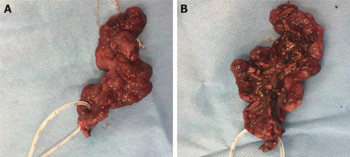
Tumours of the ampulla of Vater are relatively uncommon lesions of the digestive system. They are typically diagnosed at an earlier stage than other types of tumours in this region, due to their tendency to invoke symptoms by obstructing the bile duct or pancreatic duct. Consequently, many are potentially curable by excision. Surgical ampullectomy (SA) (or transduodenal ampullectomy) for an ampullary tumour was first described in 1899, but was soon surpassed by pancreatoduodenectomy (PD), which offered a more extensive resection resulting in a lower risk of recurrence. Ongoing innovation in endoscopic techniques over recent decades has led to the popularization of endoscopic papillectomy (EP), particularly for adenomas and even early cancers. The vast majority of resectable ampullary tumours are now treated using either PD or EP. However, SA continues to play a role in specific circumstances. Many authors have suggested specific indications for SA based on their own data, practices, or interpretations of the literature. However, certain issues have attracted controversy, such as its use for early ampullary cancers. Consequently, there has been a lack of clarity regarding indications for SA, and no evidence-based consensus guidelines have been produced. All studies reporting SA have employed observational designs, and have been heterogeneous in their methodologies. Accordingly, characteristics of patients and their tumours have differed substantially across treatment groups. Therefore, meaningful comparisons of clinical outcomes between SA, PD and EP have been elusive. Nevertheless, it appears that suitably selected cases of ampullary tumours subjected to SA may benefit from favourable peri-operative and long-term outcomes with very low mortality and significantly long survival, hence its role in this setting warrants further clarification, while it can also be useful in the management of specific benign entities. Whilst the commissioning of a randomised controlled trial seems unlikely, well-designed observational studies incorporating adjustments for confounding variables may become the best available comparative evidence for SA, potentially informing the eventual development of consensus guidelines. In this comprehensive review, we explore the role of SA in the modern management of ampullary lesions.
Keywords: Ampulla of Vater; Ampullary tumours; Endoscopic papillectomy; Pancreatoduodenectomy; Surgical ampullectomy; Transduodenal ampullectomy.
©The Author(s) 2021. Published by Baishideng Publishing Group Inc. All rights reserved.
Conflict of interest statement
Conflict-of-interest statement: The authors declare no conflict of interests for this article.
Figures
References
-
- Moekotte AL, Lof S, Van Roessel S, Fontana M, Dreyer S, Shablak A, Casciani F, Mavroeidis VK, Robinson S, Khalil K, Gradinariu G, Mowbray N, Al-Sarireh B, Fusai GK, Roberts K, White S, Soonawalla Z, Jamieson NB, Salvia R, Besselink MG, Abu Hilal M. Histopathologic Predictors of Survival and Recurrence in Resected Ampullary Adenocarcinoma: International Multicenter Cohort Study. Ann Surg. 2020;272:1086–1093. - PubMed
-
- Chareton B, Coiffic J, Landen S, Bardaxoglou E, Campion JP, Launois B. Diagnosis and therapy for ampullary tumors: 63 cases. World J Surg. 1996;20:707–712. - PubMed
-
- Moekotte AL, van Roessel S, Malleo G, Rajak R, Ecker BL, Fontana M, Han HS, Rabie M, Roberts KJ, Khalil K, White SA, Robinson S, Halimi A, Zarantonello L, Fusai GK, Gradinariu G, Alseidi A, Bonds M, Dreyer S, Jamieson NB, Mowbray N, Al-Sarireh B, Mavroeidis VK, Soonawalla Z, Napoli N, Boggi U, Kent TS, Fisher WE, Tang CN, Bolm L, House MG, Dillhoff ME, Behrman SW, Nakamura M, Ball CG, Berger AC, Christein JD, Zureikat AH, Salem RR, Vollmer CM, Salvia R, Besselink MG, Abu Hilal M International Study Group on Ampullary Cancer (ISGACA) Collaborators, Aljarrah R, Barrows C, Cagigas MN, Lai ECH, Wellner U, Aversa J, Dickson PV, Ohtsuka T, Dixon E, Zheng R, Kowalski S, Freedman-Weiss M. Development and external validation of a prediction model for survival in patients with resected ampullary adenocarcinoma. Eur J Surg Oncol. 2020;46:1717–1726. - PubMed
-
- Stolte M, Pscherer C. Adenoma-carcinoma sequence in the papilla of Vater. Scand J Gastroenterol. 1996;31:376–382. - PubMed
Publication types
LinkOut - more resources
Full Text Sources
