

A new approach for quantifying epithelial and stromal thickness changes after orthokeratology contact lens wear
- PMID: 34950488
- PMCID: PMC8692962
- DOI: 10.1098/rsos.211108
A new approach for quantifying epithelial and stromal thickness changes after orthokeratology contact lens wear
Abstract
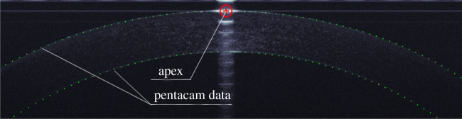
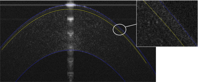
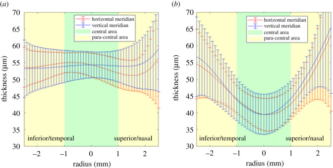
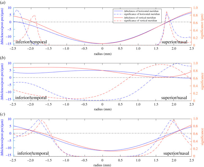
The aim of the study was to develop an automatic segmentation approach to optical coherence tomography (OCT) images and to investigate the changes in epithelial and stromal thickness profile and radius of curvature after the use of orthokeratology (Ortho-K) contact lenses. A total of 45 right eyes from 52 participants were monitored before, and after one month of, uninterrupted overnight Ortho-K lens wear. The tomography of their right eyes was obtained using optical OCT and rotating Scheimpflug imaging (OCULUS Pentacam). A custom-built MATLAB code for automatic segmentation of corneal OCT images was created and used to assess changes in epithelial thickness, stromal thickness, corneal and stromal profiles and radii of curvature before, and after one month of, uninterrupted overnight wear of Ortho-K lenses. In the central area (0-2 mm diameter), the epithelium thinned by 12.8 ± 6.0 µm (23.8% on average, p < 0.01) after one month of Ortho-K lens wear. In the paracentral area (2-5 mm diameter), the epithelium thinned nasally and temporally (by 2.4 ± 5.9 µm, 4.5% on average, p = 0.031). The stroma thickness increased in the central area (by 4.8 ± 16.1 µm, p = 0.005). The radius of curvature of the central corneal anterior surface increased by 0.24 ± 0.26 mm (3.1%, p < 0.01) along the horizontal meridian and by 0.34 ± 0.18 mm (4.2%, p < 0.01) along the vertical meridian. There were no significant changes in the anterior and posterior stromal radius of curvature. This study introduced a new method to automatically detect the anterior corneal surface, the epithelial posterior surface and the posterior corneal surface in OCT scans. Overnight wear of Ortho-K lenses caused thinning of the central corneal epithelium. The anterior corneal surface became flattered while the anterior and posterior surfaces of the stroma did not undergo significant changes. The results are consistent with the changes reported in previous studies. The reduction in myopic refractive error caused by Ortho-K lens wear was mainly due to changes in corneal epithelium thickness profile.
Keywords: OCT; contact lenses; cornea; epithelium; eye; orthokeratology.
© 2021 The Authors.
Figures
Similar articles
-
Vertical and horizontal thickness profiles of the corneal epithelium and Bowman's layer after orthokeratology.Invest Ophthalmol Vis Sci. 2013 Jan 23;54(1):691-6. doi: 10.1167/iovs.12-10263. Invest Ophthalmol Vis Sci. 2013. PMID: 23221070
-
Corneal thickness changes in myopic children during and after short-term orthokeratology lens wear.Ophthalmic Physiol Opt. 2021 Jul;41(4):757-767. doi: 10.1111/opo.12824. Epub 2021 Apr 20. Ophthalmic Physiol Opt. 2021. PMID: 33878198
-
Corneal refractive therapy with different lens materials, part 1: corneal, stromal, and epithelial thickness changes.Optom Vis Sci. 2007 Apr;84(4):343-8. doi: 10.1097/OPX.0b013e318042af1d. Optom Vis Sci. 2007. PMID: 17435518 Clinical Trial.
-
[A review of mathematical descriptors of corneal asphericity].J Fr Ophtalmol. 2002 Jan;25(1):81-90. J Fr Ophtalmol. 2002. PMID: 11965125 Review. French.
-
OCT applications in contact lens fitting.Cont Lens Anterior Eye. 2022 Aug;45(4):101540. doi: 10.1016/j.clae.2021.101540. Epub 2021 Nov 17. Cont Lens Anterior Eye. 2022. PMID: 34799247 Review.
Cited by
-
Corneal Epithelial Thickness Mapping: A Major Review.J Ophthalmol. 2024 Jan 2;2024:6674747. doi: 10.1155/2024/6674747. eCollection 2024. J Ophthalmol. 2024. PMID: 38205099 Free PMC article. Review.
-
FEA-Based Stress-Strain Barometers as Forecasters for Corneal Refractive Power Change in Orthokeratology.Bioengineering (Basel). 2024 Feb 9;11(2):166. doi: 10.3390/bioengineering11020166. Bioengineering (Basel). 2024. PMID: 38391654 Free PMC article.
-
Short-term Changes in Epithelial and Optical Redistribution Induced by Different Orthokeratology Designs.Eye Contact Lens. 2023 Dec 1;49(12):528-534. doi: 10.1097/ICL.0000000000001045. Epub 2023 Oct 30. Eye Contact Lens. 2023. PMID: 37902624 Free PMC article.
-
Long-Term Efficacy of CRT, Lucid, Euclid, and IBright Orthokeratology Lenses in Controlling Myopia Progression in Children and Adolescents: A 36-Month Retrospective Cohort Study.Clin Ophthalmol. 2025 Jul 23;19:2423-2430. doi: 10.2147/OPTH.S526689. eCollection 2025. Clin Ophthalmol. 2025. PMID: 40718507 Free PMC article.
References
-
- Mountford J. 1997. An analysis of the changes in corneal shape and refractive error induced by accelerated orthokeratology. Int. Contact Lens Clin. 24, 128-144. (10.1016/S0892-8967(97)00055-2) - DOI
LinkOut - more resources
Full Text Sources
