. 2021 Dec 23;33(1):6.
doi: 10.1007/s10856-021-06632-5.

Enhanced model protein adsorption of nanoparticulate hydroxyapatite thin films on silk sericin and fibroin surfaces

Affiliations

Enhanced model protein adsorption of nanoparticulate hydroxyapatite thin films on silk sericin and fibroin surfaces

Selçuk Özcan et al. J Mater Sci Mater Med. .

Abstract

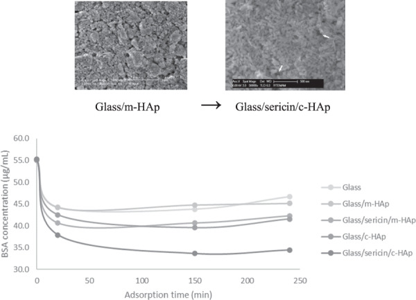
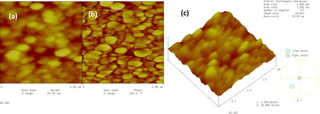
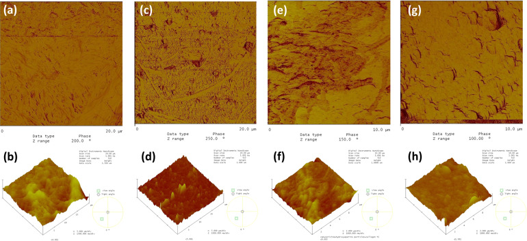
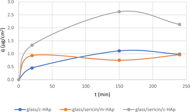
Hydroxyapatite coated metallic implants favorably combine the required biocompatibility with the mechanical properties. As an alternative to the industrial coating method of plasma spraying with inherently potential deleterious effects, sol-gel methods have attracted much attention. In this study, the effects of intermediate silk fibroin and silk sericin layers on the protein adsorption capacity of hydroxyapatite films formed by a particulate sol-gel method were determined experimentally. The preparation of the layered silk protein/hydroxyapatite structures on glass substrates, and the effects of the underlying silk proteins on the topography of the hydroxyapatite coatings were described. The topography of the hydroxyapatite layer fabricated on the silk sericin was such that the hydroxyapatite particles were oriented forming an oriented crystalline surface. The model protein (bovine serum albumin) adsorption increased to 2.62 µg/cm2 on the latter surface as compared to 1.37 µg/cm2 of hydroxyapatite on glass without an intermediate silk sericin layer. The BSA adsorption on glass (blank), glass/c-HAp, glass/m-HAp, glass/sericin/c-HAp, and glass/sericin/m-HAp substrates, reported as decrease in BSA concentration versus contact time.


Conflict of interest statement

The authors declare no competing interests.

Figures

None
The BSA adsorption on glass (blank), glass/c-HAp, glass/m-HAp, glass/sericin/c-HAp, and glass/sericin/m-HAp substrates, reported as decrease in BSA concentration versus contact time.
Fig. 1
Fig. 1
The SEM images of the c-HAp coatings on the glass substrates prepared by 15w% solid content sol by dip coating, at various magnifications (a) 500x, (b) 50,000x, and the m-HAp coatings on the glass substrates prepared by 15w% solid content sol by dip coating at various magnifications (c) 500x, (d) 50,000x.
Fig. 2
Fig. 2
The SEM images of the thin films prepared with 15w% HAp suspensions after the heat treatment at 560 °C, for 30 min. with a heating rate of 15 °C/min at various magnifications (a) c-HAp 50,000x, (b) m-HAp 50,000x.
Fig. 3
Fig. 3
The AFM images of the sericin film on glass substrate, (a) 2-dim. topographical, (b) phase, and (c) 3-dim. topographical images, 1.84 × 1.84 μm.
Fig. 4
Fig. 4
The AFM images of the fibroin film on glass substrate, (a) 2-dim. topographical, (b) phase, and (c) 3-dim. topographical images, 1.84 × 1.84 μm.
Fig. 5
Fig. 5
The SEM and AFM images of the c-HAp and m-HAp films (with 15w% solid content suspension) on the glass/sericin substrate (a) the SEM image of c-HAp film, 65,000x, (b) the AFM 3-dim. topographical image of c-HAp film, 10 × 10 μm, (c) the SEM image of m-HAp film, 50,000x (d) the AFM 3-dim. topographical image of m-HAp film, 10 × 10 μm.
Fig. 6
Fig. 6
The SEM and AFM images of the c-HAp and m-HAp films (with 15w% solid content suspension) on the glass/fibroin substrate (a) the SEM image of c-HAp film, 50,000x, (b) the AFM 3-dim. topographical image of c-HAp film, 10 × 10 μm, (c) the SEM image of m-HAp film, 35,000x (d) the AFM 3-dim. topographical image of m-HAp film, 10 × 10 μm.
Fig. 7
Fig. 7
SEM images of (a) the glass/sericin/c-HAp, (b) the glass/sericin/m-HAp, (c) the glass/fibroin/c-HAp, (d) the glass/fibroin/m -HAp thin film structures after heat treatment at 560 °C; the arrows indicate some of the hole-like gaps through which sericin and fibroin burnt out.
Fig. 8
Fig. 8
The AFM phase images of adsorbed BSA on (a) glass, (b) glass/c-HAp film (c) glass/m-HAp film.
Fig. 9
Fig. 9
The AFM images of adsorbed BSA on (a) the glass/fibroin/c-HAp; phase image, (b) the glass/fibroin/c-HAp; 3-dim. topographical image, (c) the glass/fibroin/m-HAp; phase image, (d) the glass/fibroin/m-HAp; 3-dim. topographical image, (e) the glass/sericin/c-HAp; phase image, (f) the glass/sericin/c-HAp; 3-dim. topographical image, (g) the glass/sericin/m-HAp films; phase image, (h) the glass/sericin/m-HAp films; 3-dim. topographical image.
Fig. 10
Fig. 10
The AFM phase image of adsorbed collagen type I on (a) the glass, (b) the glass/c-HAp film, (c) the glass/m-HAp film.
Fig. 11
Fig. 11
The AFM images of adsorbed collagen type I on (a). a the glass/fibroin/c-HAp; phase image, (b) the glass/fibroin/c-HAp; 3-dim. topographical image, (c) the glass/fibroin/m-HAp; phase image, (d) the glass/fibroin/m-HAp; 3-dim. topographical image, (e) the glass/sericin/c-HAp; phase image, (f) the glass/sericin/c-HAp; 3-dim. topographical image, (g) the glass/sericin/m-HAp films; phase image, (h) the glass/sericin/m-HAp films; 3-dim. topographical image.
Fig. 12
Fig. 12
The BSA adsorption on glass (blank), glass/c-HAp, glass/m-HAp, glass/sericin/c-HAp, and glass/sericin/m-HAp substrates, reported as decrease in BSA concentration versus contact time.
Fig. 13
Fig. 13
BSA adsorbed quantity as a function of time on the glass/c-HAp, the glass/sericin/m-HAp, and the glass/sericin/c-HAp films.

References

    1. Guyton AC. Textbook of medical physiology. 8th ed. Philadelphia: W. B. Saunders Company; 1991.
    1. Winn SR, Uludag H, Hollinger JO. Sustained release emphasizing recombinant human bone morphogenetic protein-2. Adv Drug Deliv Rev. 1998;31:303–18. doi: 10.1016/S0169-409X(97)00126-9. - DOI - PubMed
    1. Hench LL. Bioceramics. J Am Ceram Soc. 1998;81:1705–28. doi: 10.1111/j.1151-2916.1998.tb02540.x. - DOI
    1. Albrektsson T, Johansson C. Osteoinduction, osteoconduction and osseointegration. Eur Spine J. 2001;10:S96–101. doi: 10.1007/s005860100282. - DOI - PMC - PubMed
    1. Davies JE, Hosseini MM. Histodynamics of endosseous wound healing. Davies JE. editor, Chapter 1: 1-14, Bone Engineering. Toronto: Em Squared Inc.; 2000.

MeSH terms