. 2021 Dec 24:10:e72747.
doi: 10.7554/eLife.72747.

A dual endosymbiosis supports nutritional adaptation to hematophagy in the invasive tick Hyalomma marginatum

Affiliations

A dual endosymbiosis supports nutritional adaptation to hematophagy in the invasive tick Hyalomma marginatum

Marie Buysse et al. Elife. .

Abstract

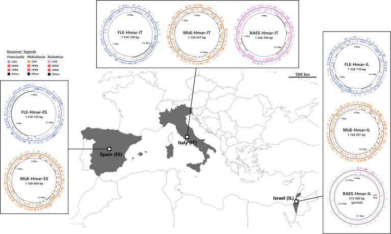
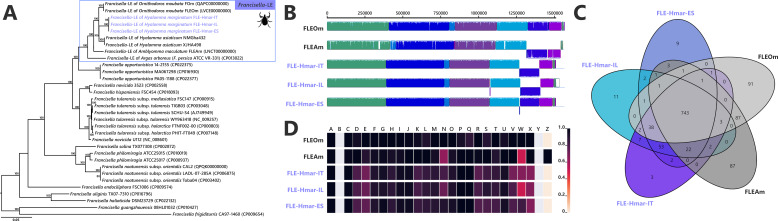
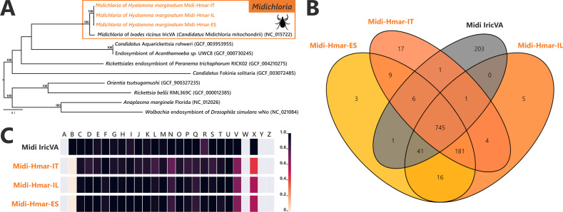
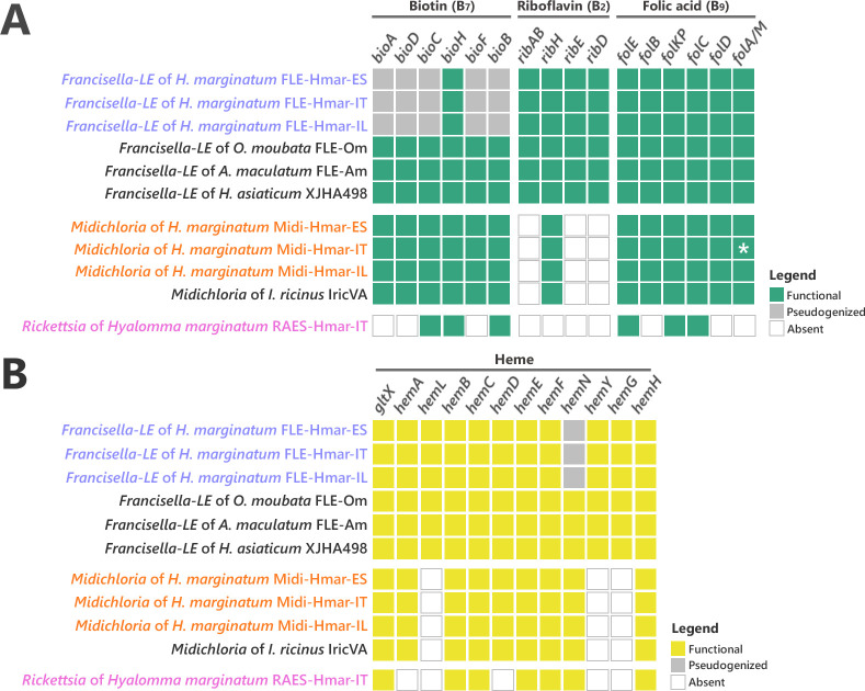
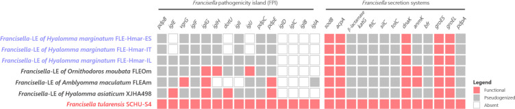
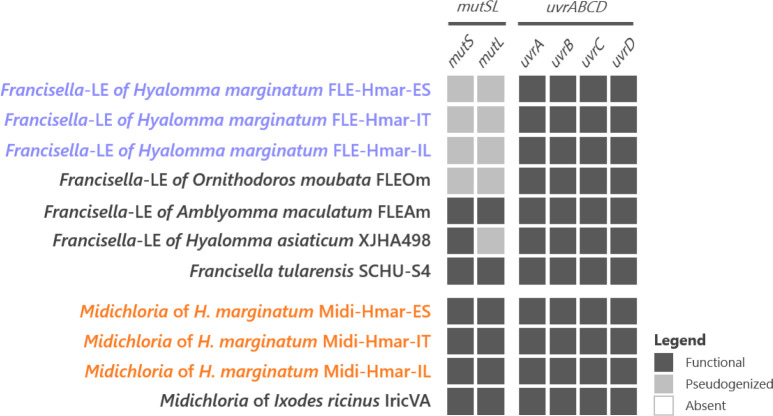
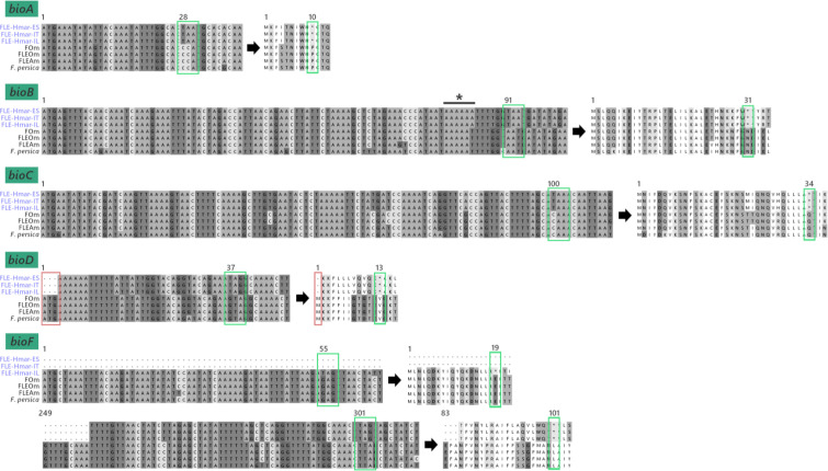
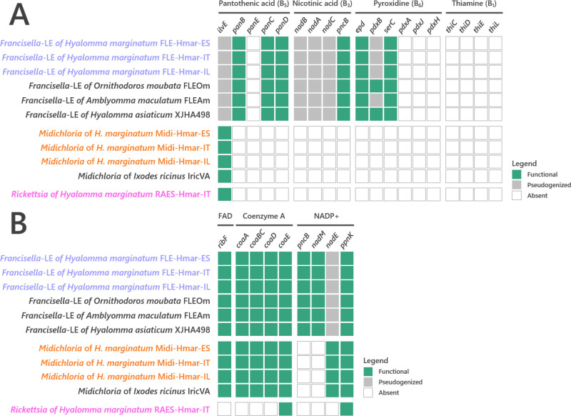
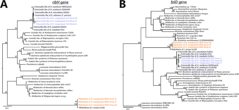
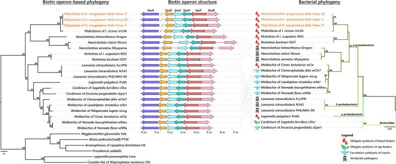
Many animals are dependent on microbial partners that provide essential nutrients lacking from their diet. Ticks, whose diet consists exclusively on vertebrate blood, rely on maternally inherited bacterial symbionts to supply B vitamins. While previously studied tick species consistently harbor a single lineage of those nutritional symbionts, we evidence here that the invasive tick Hyalomma marginatum harbors a unique dual-partner nutritional system between an ancestral symbiont, Francisella, and a more recently acquired symbiont, Midichloria. Using metagenomics, we show that Francisella exhibits extensive genome erosion that endangers the nutritional symbiotic interactions. Its genome includes folate and riboflavin biosynthesis pathways but deprived functional biotin biosynthesis on account of massive pseudogenization. Co-symbiosis compensates this deficiency since the Midichloria genome encompasses an intact biotin operon, which was primarily acquired via lateral gene transfer from unrelated intracellular bacteria commonly infecting arthropods. Thus, in H. marginatum, a mosaic of co-evolved symbionts incorporating gene combinations of distant phylogenetic origins emerged to prevent the collapse of an ancestral nutritional symbiosis. Such dual endosymbiosis was never reported in other blood feeders but was recently documented in agricultural pests feeding on plant sap, suggesting that it may be a key mechanism for advanced adaptation of arthropods to specialized diets.

Keywords: Francisella; Hyalomma; Midichloria; endosymbiosis; evolutionary biology; hematophagy.


Conflict of interest statement

MB, AF, YG, TN, FC, EO, AG, AP, BM, CB, AC, DS, OD No competing interests declared

Figures

Figure 1.
Figure 1.. Sampled locations for Hyalomma marginatum ticks.
Sampling countries are colored in gray. The symbiont genomes retrieved from each site are graphically represented in blue, orange, and pink, corresponding to Francisella-LE (FLE), Midichloria (Midi), and R. aeschlimannii (RAES), respectively. Circles on genome maps correspond to the following: (1) forward strand genes; (2) reverse strand genes; (3) in gray and black, contigs. Circular maps of the genomes were produced using CGView.
Figure 1—figure supplement 1.
Figure 1—figure supplement 1.. Rickettsia phylogenetic tree assessing the affiliation of the Rickettsia detected in Hyalomma marginatum specimens from Italy and Israel.
The phylogenetic tree was inferred using maximum likelihood from a concatenated alignment of 324 single-copy orthologs genes (88,128 amino acids; best-fit approximation for the evolutionary model: JTT+G4+I). The numbers on each node represent the support of 1000 bootstrap replicates; only bootstrap values >70% are shown. The scale bar is in units of substitution/site. Sequence from the newly sequenced Rickettsia genome is indicated by a pink font.
Figure 1—figure supplement 2.
Figure 1—figure supplement 2.. Phylogenetic relationship of the Hyalomma species used in our study.
The phylogenetic tree was inferred using maximum likelihood from an alignment of the cytochrome c oxidase I (cox1) gene sequences (546 bp unambiguously aligned, best-fit approximation for the evolutionary model: HKY+G+I). The numbers on each node represent the support of 1000 bootstrap replicates; only bootstrap values >80% are shown. The scale bar is in units of substitution/site.
Figure 1—figure supplement 3.
Figure 1—figure supplement 3.. Presence of FLE and Midichloria within the Hyalomma genus.
The Hyalomma cladogram was adapted from cox1 phylogeny showed in Figure 6. The numbers on each node represent the support of 1000 bootstrap replicates; only bootstrap values >80% are shown. The scale bar is in units of substitution/site. Blue and orange plain circles indicate the presence of FLE and Midichloria, respectively, as detailed in Supplementary file 1d. Blank circle represents an absence of symbiont.
Figure 2.
Figure 2.. Comparative genomic features of FLE.
(A) Whole-genome phylogenetic relationship of the three FLE genomes obtained from Hyalomma marginatum specimens (highlighted), FLE of other tick species, and Francisella pathogenic species, including F. tularensis. The phylogenetic tree was inferred using maximum likelihood from a concatenated alignment of 436 single-copy orthologs genes (132,718 amino acids; best-fit approximation for the evolutionary model: CPREV+G4+I). The numbers on each node represent the support of 1000 bootstrap replicates; only bootstrap values >70% are shown. The scale bar is in units of substitution/site. Sequences from newly sequenced FLE genomes are indicated by a blue font. (B) Whole-genome synteny of the three FLE genomes obtained from H. marginatum specimens (blue font) and representative FLE (black font). Each contiguously colored locally collinear block (LCB) represents a region without rearrangement of the homologous backbone sequence. LCBs below the center in Francisella genomes represent blocks in the reverse orientation. The height of color bars within LCBs corresponds to the average level of similarity (conservation) profile in that region of the genome sequence. Areas that are completely white were not aligned and containd sequence elements specific to a particular genome. (C) Venn diagram representing orthologs distribution between the three FLE genomes obtained from H. marginatum specimens (blue font) and representative FLE (black font). (D) Clusters of orthologous groups of proteins (COGs) comparisons between the three FLE genomes obtained from H. marginatum specimens (blue font) and representative FLE (black font). The color scale indicates the percentage of functional genes per category. Gray color means that genes are not present in a given category. The COG category names (capital letters from A to Z) correspond to the NCBI reference naming system.
Figure 3.
Figure 3.. Comparative genomic features of Midichloria.
(A) Whole-genome phylogenetic relationship of the three Midichloria genomes obtained from Hyalomma marginatum specimens (highlighted), 'Ca M. mitochondrii', and other Rickettsiales species. The phylogenetic tree was inferred using maximum likelihood from a concatenated alignment of 242 single-copy orthologs genes (49,957 amino acids; best-fit approximation for the evolutionary model: LG+G4+I). The numbers on each node represent the support of 1000 bootstrap replicates; only bootstrap values >70% are shown. The scale bar is in units of substitution/site. Sequences from newly sequenced Midichloria genomes are indicated by an orange font. (B) Venn diagram representing orthologs distribution between the three Midichloria genomes obtained from H. marginatum specimens (orange font) and ‘Ca. M. mitochondrii’ (black font). (C) Clusters of orthologous groups of proteins (COGs) comparisons between the three Midichloria genomes obtained from H. marginatum specimens (orange font) and ‘Ca. M. mitochondrii’ (black font). The color scale indicates the percentage of functional genes per category. Gray color means that genes are not present in a given category. The COG category names (capital letters from A to Z) correspond to the NCBI reference naming system.
Figure 4.
Figure 4.. Conservation level of biosynthetic pathways involved in the symbiotic interaction between ticks and endosymbionts.
(A) Biosynthetic pathways for the three key B vitamins (biotin, folic acid, and riboflavin) required for tick nutrition of FLE, Midichloria, and Rickettsia. Newly sequenced FLE, Midichloria, and Rickettsia genomes are indicated by a blue, an orange, and a pink font, respectively. Green squares, functional genes; gray squares, pseudogenes; white squares, missing genes; asterisk, partial gene sequence that do not contain internal codon stop but that are located at the beginning of a contig. (B) Heme biosynthetic pathway of FLE, Midichloria, and Rickettsia. Newly sequenced FLE, Midichloria, and Rickettsia genomes are indicated by a blue, an orange, and a pink font, respectively. Yellow squares, functional genes; gray squares, pseudogenes; white squares, missing genes.
Figure 4—figure supplement 1.
Figure 4—figure supplement 1.. Conservation level of the genes involved in the Francisella pathogenicity island (FPI) on FLE of Hyalomma marginatum and references genomes.
Genomes of FLE of H. marginatum are indicated by a blue font. Francisella pathogenic species is indicated by a red font. Red squares, functional genes; gray squares, pseudogenes; white squares, missing genes.
Figure 4—figure supplement 2.
Figure 4—figure supplement 2.. Conservation level of the genes involved in the mismatch repair system (mutSL) and the nucleotide excision repair system (uvrABCD) on FLE and Midichloria of Hyalomma marginatum and references genomes.
Newly sequenced FLE and Midichloria genomes are indicated by a blue and an orange font, respectively. Black squares, functional genes; gray squares, pseudogenes; white squares, missing genes.
Figure 4—figure supplement 3.
Figure 4—figure supplement 3.. Pseudogenization of biotin synthesis genes.
Representative nucleotide and amino sequences are shown for each of the five pseudogenized biotin genes. Green box, premature stop codon; red box, absence of start codon; asterisk, insertion of one adenine base on a poly(A) zone and inducing a frameshift in the bioB coding sequence. Numbers attached to the boxes correspond to the position of the first nucleic acid leading to the premature stop codon and its position in the protein sequence.
Figure 4—figure supplement 4.
Figure 4—figure supplement 4.. Conservation level of (A) pantothenic acid, nicotinic acid, pyroxidine, and thiamine B vitamins and (B) cofactors biosynthetic pathways of FLE, Midichloria, and Rickettsia.
Newly sequenced FLE, Midichloria, and Rickettsia genomes are indicated by a blue, an orange, and a pink font, respectively. Green squares, functional genes; gray squares, pseudogenes; white squares, missing genes.
Figure 4—figure supplement 5.
Figure 4—figure supplement 5.. Phylogenetic trees of representative genes of riboflavin and folate biosynthesis pathways of FLE and Midichloria.
The phylogenetic trees of (A) ribH (riboflavin pathway), and (B) folD (folate) were inferred using maximum likelihood from amino acid alignments. The numbers on each node represent the support of 1000 bootstrap replicates; only bootstrap values >70% are shown. The scale bar is in units of substitution/site. ribH: 129 amino acids unambiguously aligned, best-fit approximation for the evolutionary model LG+G4; folD: 273 amino acids unambiguously aligned, best-fit approximation for the evolutionary model CPREV+G4+I. Newly sequenced FLE and Midichloria sequences are indicated by a blue and an orange font, respectively, while sequences from other FLE and Midichloria are highlighted in blue and orange, respectively.
Figure 5.
Figure 5.. Evolutionary relationships (left) and structure (middle) of the streamlined biotin operon, confronted to the bacterial phylogeny (right).
The phylogenetic tree of the streamlined biotin operon (left) was inferred using maximum likelihood (ML) from a concatenated alignment of the six genes composing the biotin biosynthetic pathway (1493 amino acids; best-fit approximation for the evolutionary model: CPREV+G4+I). The numbers on each node represent the support of 1000 bootstrap replicates; only bootstrap values >70% are shown. The scale bar is in units of substitution/site. Sequences from newly sequenced Midichloria genomes are indicated by an orange font. Structure of the streamlined biotin operon (middle) is represented by arrows in which each colored arrow corresponds to an intact gene and its direction. A discontinuous line corresponds to genes encoding on two separated contigs. The bacterial phylogenetic tree (right) was inferred using ML from an alignment of 16S rDNA sequences (1362 bp unambiguously aligned; best-fit approximation for the evolutionary model: GTR+G+I). The numbers on each node represent the support of 1000 bootstrap replicates; only bootstrap values >70% are shown. The scale bar is in units of substitution/site. Sequences from Midichloria of Hyalomma marginatum are indicated by an orange font.
Figure 6.
Figure 6.. FLE and Midichloria loads in ovaries of Hyalomma marginatum.
(A) Representation and statistical comparison of FLE and Midichloria densities. (B) FLE density as a function of Midichloria density in H. marginatum ovaries. Gray area represents 95% confidence interval.

References

    1. Akhter S, Aziz RK, Edwards RA. PhiSpy: a novel algorithm for finding prophages in bacterial genomes that combines similarity- and composition-based strategies. Nucleic Acids Research. 2012;40:1–13. doi: 10.1093/nar/gks406. - DOI - PMC - PubMed
    1. Alonge M, Soyk S, Ramakrishnan S, Wang X, Goodwin S, Sedlazeck FJ, Lippman ZB, Schatz MC. RaGOO: fast and accurate reference-guided scaffolding of draft genomes. Genome Biology. 2019;20:1–17. doi: 10.1186/s13059-019-1829-6. - DOI - PMC - PubMed
    1. Andrews S. FastQC: A Quality Control Tool for High Throughput Sequence Data. ScienceOpen, Inc; 2010.
    1. Azagi T, Klement E, Perlman G, Lustig Y, Mumcuoglu KY, Apanaskevich DA, Gottlieb Y. Francisella-Like Endosymbionts and Rickettsia Species in Local and Imported Hyalomma Ticks. Applied and Environmental Microbiology. 2017;83:1–14. doi: 10.1128/AEM.01302-17. - DOI - PMC - PubMed
    1. Bankevich A, Nurk S, Antipov D, Gurevich AA, Dvorkin M, Kulikov AS, Lesin VM, Nikolenko SI, Pham S, Prjibelski AD, Pyshkin AV, Sirotkin AV, Vyahhi N, Tesler G, Alekseyev MA, Pevzner PA. SPAdes: a new genome assembly algorithm and its applications to single-cell sequencing. Journal of Computational Biology. 2012;19:455–477. doi: 10.1089/cmb.2012.0021. - DOI - PMC - PubMed

Publication types

Substances