. 2022 Jun 1;24(6):888-900.
doi: 10.1093/neuonc/noab292.

ATRX loss promotes immunosuppressive mechanisms in IDH1 mutant glioma

Affiliations

ATRX loss promotes immunosuppressive mechanisms in IDH1 mutant glioma

Chengchen Hu et al. Neuro Oncol. .

Abstract

Background: ATRX inactivation occurs with IDH1R132H and p53 mutations in over 80% of Grades II/III astrocytomas. It is believed that ATRX loss contributes to oncogenesis by dysregulating epigenetic and telomere mechanisms but effects on anti-glioma immunity have not been explored. This paper examines how ATRX loss contributes to the malignant and immunosuppressive phenotypes of IDH1R132H/p53mut glioma cells and xenografts.

Methods: Isogenic astrocytoma cells (+/-IDH1R132H/+/-ATRXloss) were established in p53mut astrocytoma cell lines using lentivirus encoding doxycycline-inducible IDH1R132H, ATRX shRNA, or Lenti-CRISPR/Cas9 ATRX. Effects of IDH1R132H+/-ATRXloss on cell migration, growth, DNA repair, and tumorigenicity were evaluated by clonal growth, transwell and scratch assays, MTT, immunofluorence and immunoblotting assays, and xenograft growth. Effects on the expression and function of modulators of the immune microenvironment were quantified by qRT-PCR, immunoblot, T-cell function, macrophage polarization, and flow cytometry assays. Pharmacologic inhibitors were used to examine epigenetic drivers of the immunosuppressive transcriptome of IDH1R132H/p53mut/ATRXloss cells.

Results: Adding ATRX loss to the IDH1R132H/p53mut background promoted astrocytoma cell aggressiveness, induced expression of BET proteins BRD3/4 and an immune-suppressive transcriptome consisting of up-regulated immune checkpoints (e.g., PD-L1, PD-L2) and altered cytokine/chemokine profiles (e.g., IL33, CXCL8, CSF2, IL6, CXCL9). ATRX loss enhanced the capacity of IDH1R132H/p53mut cells to induce T-cell apoptosis, tumorigenic/anti-inflammatory macrophage polarization and Treg infiltration. The transcriptional and biological immune-suppressive responses to ATRX loss were enhanced by temozolomide and radiation and abrogated by pharmacologic BET inhibition.

Conclusions: ATRX loss activates a BRD-dependent immune-suppressive transcriptome and immune escape mechanism in IDH1R132H/p53mut astrocytoma cells.

Keywords: ATRX loss; BRD-dependent immune-suppressive transcriptome; IDH1 mutant astrocytoma; glioma immune microenvironment.


Figures

Fig. 1
Fig. 1
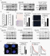
ATRX loss promotes aggressive phenotypes in IDH1R132H/p53mut astrocytoma cells. P53 mutant U373 and Snb19 cells +/−IDH1R132H, +/−ATRXloss were established using lentivirus encoding either doxycycline (DOX) inducible IDH1R132H or shRNA against ATRX (or control) as described in Materials and Methods. A. Immunoblots showing induction of IDH1R132H by DOX and ATRX expression inhibition by sh-ATRX and CRISPR/Cas9 ATRX. B. 2-Hydroxyglutarate (2-HG) levels in conditioned medium of U373 and Snb19 cells expressing +/−IDH1R132H measured by mass spectrometry. C. Clonogenicity of U373 cells (IDH1R132H/p53mut +/−ATRXloss) in soft agar. Images of colony densities are shown (left panel). Colonies >100µm diameter were quantified by computer-assisted image analysis (right panel). D. Invasion capacities of U373 IDH1R132H/p53mut+/−ATRXloss cells measured by transwell invasion assay. E. Immunoblot showing effects of TMZ, γ-radiation or TZP on γH2AX expression in U373 IDH1R132H/p53mut+/−ATRXloss cells. F. Immunofluorescent staining and quantification of γH2AX expression in IDH1R132H/p53mut/ATRXloss cells treated +/−TMZ. G. Viability measured by MTT assay of U373 IDH1R132H/p53mut+/−ATRXloss treated with TMZ or PARP inhibitor TZP for 72 h. *P < .05, **P < .01, and ***P < .001.
Fig. 2
Fig. 2
ATRX loss induces PD-L1 and an immunosuppressive cytokine/chemokine profile in IDH1R132H/p53mut cells in vitro and in vivo. A. Expression quantification by qRT-PCR of 22 immune checkpoint ligands in IDH1R132H/p53mut+/−ATRXloss U373 cells; inhibitory immune checkpoint molecules are labeled red and co-stimulatory ligand is labeled green. B. Immunoblots showing PD-L1 expression in IDH1R132H/p53mut/ATRXloss, IDH1R132H/p53mut, ATRXloss/p53mut, and p53mut U373 and Snb19 cells. C. Immunofluorescence showing membrane distribution of PD-L1 in IDH1R132H/p53mut+/−ATRXloss U373 cells. D. Expression quantification by qRT-PCR of 21 cytokines and chemokines in IDH1R132H/p53mut+/−ATRXloss U373 cells (left panel). E. Immunoblots showing IL33 level in whole cell extracts. F. and G. Immunohistochemistry and quantification of PD-L1 and IL33 in IDH1R132H/p53mut+/−ATRXloss U373 tumor xenografts. H. IL-33 and CXCL3 expression in TCGA IDH1mut/non-1p19q co-deleted ATRX-mutant vs. ATRX-WT clinical samples (http://gliovis.bioinfo.cnio.es). *P < .05, **P < .01, and ***P < .001.
Fig. 3
Fig. 3
Chemo-radiation augments the immunosuppressive transcriptome of IDH1R132H/p53mut/ATRXloss astrocytoma cells. A. and B. PD-L1 expression in IDH1R132H/p53mut+/−ATRXloss U373 and Snb19 cells treated with TMZ (600 μM) and radiation (5 Gy) for 5 h (qRT-PCR, panel A) and for 48 h (immunoblot, panel B). C. Cell-surface PD-L1 expression determined by flow cytometry in patient-derived IDH1R132H/p53mut/ATRXloss MGG119 and MGG152 astrocytoma cells treated with TMZ (600 μM) and radiation (5Gy) for 48 h. D. qRT-PCR analysis measuring expression of key cytokines/chemokines in IDH1R132H/p53mut+/-ATRXloss U373 cells treated with TMZ or IR. E. qRT-PCR analysis measuring expression of key cytokines/chemokines in IDH1R132H/p53mut/ATRXloss MGG119 cells treated with TMZ and radiation. *P < .05, **P < .01, and ***P < .001.
Fig. 4
Fig. 4
ATRX loss in IDH1 mutant glioma cells suppresses T cells and induces immune-suppressive M2 macrophages. A. IDH1R132H/p53mut+/-ATRXloss U373 cells were co-cultured with activated CD8+ T cells (10:1) and CD8 T-cell apoptosis measured by caspase 3/7 activation. B. IDH1R132H/p53mut+/−ATRXloss U373 cells were co-cultured with activated CD8+ T cells (1:5) for 24 h, remaining viable adherent cells shown in phase-contrast photomicrographs (left panel) and quantified by CCK-8 assay (right panel). C. IDH1R132H/p53mut+/−ATRXloss U373 cells were treated +/−TMZ for 48 h prior to culture with activated Jurkat cells +/−anti-PD-L1 antibody and Jurkat cell apoptosis measured by caspase 3/7 activation (left panel). IL2 production as a marker of Jurkat cell activation was measured by ELISA (right panel). D. Activated, CFSE-labeled PBMC-derived CD8+ T cells were co-cultured with macrophages pretreated with conditioned medium from ATRXwt or ATRXloss cells and T-cell proliferation quantified by flow cytometry (left panel). Histogram shows CD8+ T-cell numbers (right panel). E. Expression levels of CD8A and CD68 in clinical ATRXmut vs. ATRXwt TCGA-IDHmut-non-1p19q co-deletion astrocytoma datasets (http://gliovis.bioinfo.cnio.es). F. TIMER datasets showing infiltration of CD8+ T cells and macrophages in ATRXmut vs. ATRXwt glioma samples (http://cistrome.org/TIMER). *P < .05, **P < .01, and ***P < .001.
Fig. 5
Fig. 5
ATRX loss promotes an immunosuppressive microenvironment in an immune-competent model of orthotopic astrocytoma. A. Immunoblots showing induction of IDHR132H by DOX, loss of ATRX expression by sh-ATRX and PD-L1 induction in murine GL261 astrocytoma cells. B. qRT-PCR analysis measuring expression of six cytokines and chemokines in IDH1R132H/p53mut+/−ATRXloss GL261 cells. C. Representative H&E staining and size quantification of xenografts derived from IDH1R132H/p53mut+/−ATRXloss GL261 cells. D. and E. Immunofluorescence staining and quantification of Iba-1+ (green), M2 marker Arg1+ (red) and PD-L1(red) in orthotopic tumor xenografts derived from IDH1R132H/p53mut+/−ATRXloss GL261 cells. F. Immunohistochemistry staining and quantification of FOXP3+ T cells (brown) in orthotopic tumor xenografts derived from IDH1R132H/p53mut+/−ATRXloss GL261 cells (n = 8 random fields per group). *P < .05, **P < .01, ***P < .001. See online supplementary material for a color version of this figure.
Fig. 6
Fig. 6
Pan-BET bromodomain inhibitors attenuate immunosuppressive transcriptome and immunosuppressive responses to IDH1R132H/p53mut/ATRXloss. A. qRT-PCR quantification of PD-L1 expression in IDH1R132H/p53mut/ATRXloss glioma cells treated with the indicated epigenetic inhibitors for 72 h. B. immunoblot analysis of PD-L1 expression in IDH1R132H/p53mut+/−ATRXloss glioma cells treated with JQ1(JQ+) or inactive control (JQ−) for 72 h; C. qRT-PCR quantification of key cytokines/chemokines in IDH1R132H/p53mut/ATRXloss glioma cells treated with JQ1(+/−) for 72 h. D. qRT-PCR quantification of BRDs-1,2,3,4 in IDH1R132H/p53mut+/−ATRXloss glioma cells. E. Immunoblot of BRD3 expression in +/−IDH1R132H/p53mut+/−ATRXloss U373 and SNB19 glioma cells. F. BRD3 expression levels in clinical Grades II, III and IV TCGA glioma datasets (left panel) and in IDHmut/non-1p19q co-deleted ATRX wild-type and ATRX-mutant TCGA glioma datasets (right panel) (http://gliovis.bioinfo.cnio.es/). G. Model of immune escape mechanisms promoted by ATRX loss in IDH1R132H/p53mut astrocytoma. *P < .05, **P < .01, and ***P < .001.

Comment in

References

    1. Claus EB, Walsh KM, Wiencke JK, et al. . Survival and low-grade glioma: the emergence of genetic information. Neurosurg Focus. 2015;38(1):E6. - PMC - PubMed
    1. Brat DJ, Verhaak RG, et al. ; Cancer Genome Atlas Research Network. . Comprehensive, integrative genomic analysis of diffuse lower-grade gliomas. N Engl J Med. 2015;372(26):2481–2498. - PMC - PubMed
    1. Johnson BE, Mazor T, Hong C, et al. . Mutational analysis reveals the origin and therapy-driven evolution of recurrent glioma. Science. 2014;343(6167):189–193. - PMC - PubMed
    1. Mukherjee J, Johannessen TC, Ohba S, et al. . Mutant IDH1 cooperates with ATRX loss to drive the alternative lengthening of telomere phenotype in glioma. Cancer Res. 2018;78(11):2966–2977. - PMC - PubMed
    1. Nunez FJ, Mendez FM, Kadiyala P, et al. . IDH1-R132H acts as a tumor suppressor in glioma via epigenetic up-regulation of the DNA damage response. Sci Transl Med. 2019;11(479). - PMC - PubMed

Publication types