. 2021 Dec 24;11(1):24430.
doi: 10.1038/s41598-021-04047-4.

Reporter gene-based optoacoustic imaging of E. coli targeted colon cancer in vivo

Affiliations

Reporter gene-based optoacoustic imaging of E. coli targeted colon cancer in vivo

Misun Yun et al. Sci Rep. .

Abstract

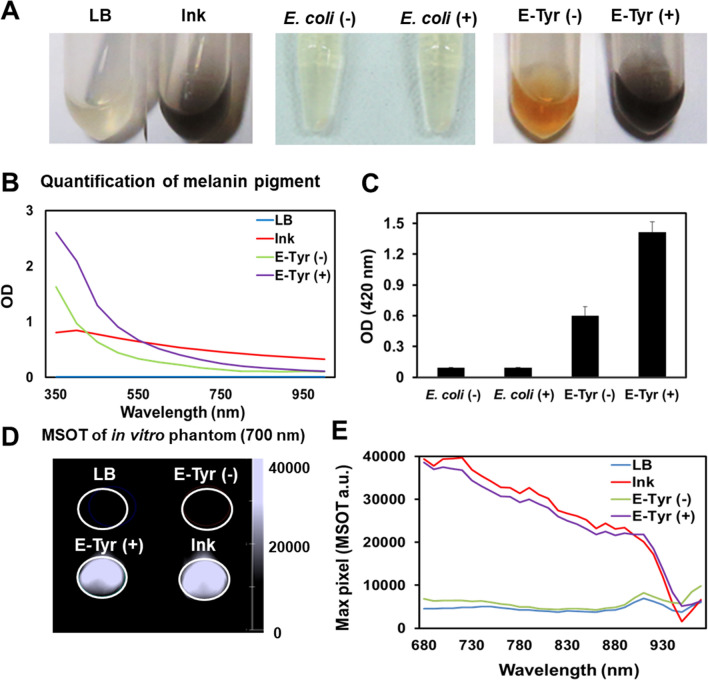
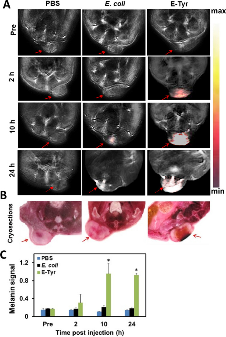
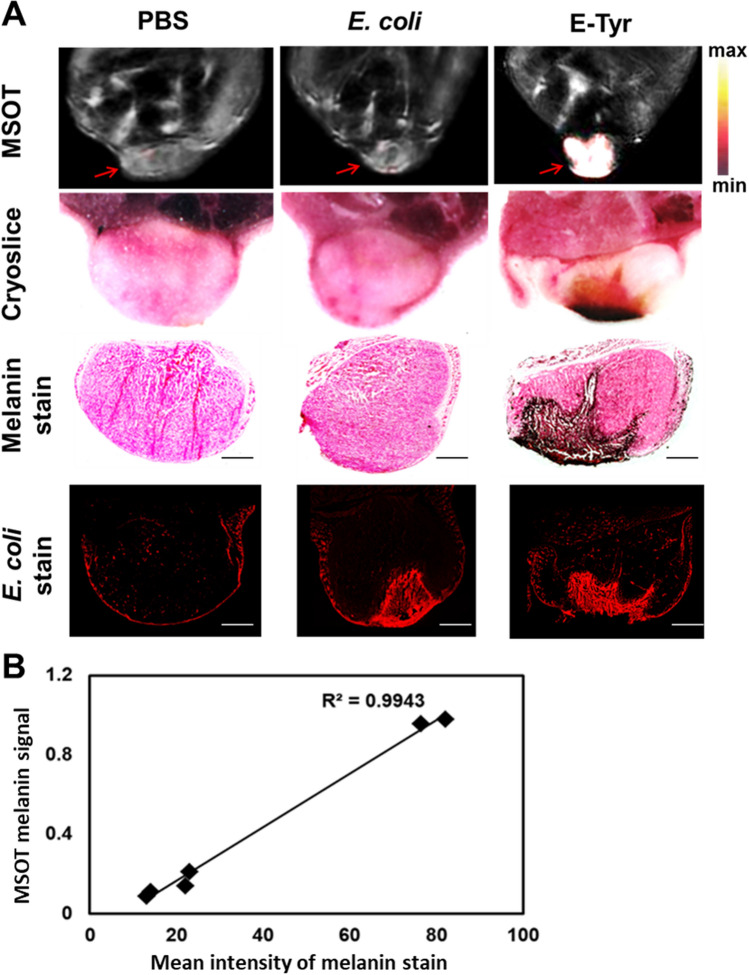
Bacteria-mediated cancer-targeted therapy is a novel experimental strategy for the treatment of cancers. Bacteria can be engineered to overcome a major challenge of existing therapeutics by differentiating between malignant and healthy tissue. A prerequisite for further development and study of engineered bacteria is a suitable imaging concept which allows bacterial visualization in tissue and monitoring bacterial targeting and proliferation. Optoacoustics (OA) is an evolving technology allowing whole-tumor imaging and thereby direct observation of bacterial colonization in tumor regions. However, bacterial detection using OA is currently hampered by the lack of endogenous contrast or suitable transgene fluorescent labels. Here, we demonstrate improved visualization of cancer-targeting bacteria using OA imaging and E. coli engineered to express tyrosinase, which uses L-tyrosine as the substrate to produce the strong optoacoustic probe melanin in the tumor microenvironment. Tumors of animals injected with tyrosinase-expressing E. coli showed strong melanin signals, allowing to resolve bacterial growth in the tumor over time using multispectral OA tomography (MSOT). MSOT imaging of melanin accumulation in tumors was confirmed by melanin and E. coli staining. Our results demonstrate that using tyrosinase-expressing E. coli enables non-invasive, longitudinal monitoring of bacterial targeting and proliferation in cancer using MSOT.


Conflict of interest statement

V.N. is a shareholder at iThera Medical GmbH, Munich, Germany. The remaining authors declare no competing interests.

Figures

Figure 1
Figure 1
Production of melanin by E-Tyr in vitro. (A) Melanin production in vitro. Tyrosinase-expressing E. coli (E-Tyr) were grown overnight in the presence or absence of L-tyrosine supplementation. (left) LB (growth medium) and ink control, (middle) supernatant of E. coli with the tyrosinase-expression vector (control bacteria) cultured with (E. coli(+)) or without (E. coli(−)) L-tyrosine supplementation, and (right) supernatant of E. coli carrying the tyrosinase-expression vector cultured with (E-Tyr(+)) or without (E-Tyr(−)) l-tyrosine supplementation. (B) Optical density measurements of LB, ink, E-Tyr (−) and E-Tyr (+) over the spectrum from 350 to 1000 nm. (C) Quantification of melanin pigment shown in (A), E. coli (−), E. coli (+), E-Tyr (−) and E-Tyr (+), using a spectrophotometer at 420 nm wavelength. (D) Optoacoustic in vitro phantom imaging of (C) with an optoacoustic image spectrum peaked at 700 nm. (E) Optoacoustic spectra for E-Tyr with or without L-tyrosine supplementation, ink and LB media, measured between 680 and 970 nm.
Figure 2
Figure 2
MSOT imaging of CT26 tumors in mice after injection with E-Tyr. (A) MSOT images of melanin signal in CT26 tumor bearing live mice (n = 6/group) with the indicated time after E-Tyr (1 × 108 cfu) injection. Unmixed melanin overlaid on anatomy. The tumor area is indicated by a red arrow (left; PBS injected control mice, middle; E. coli injected mice, right; E-Tyr injected mice) (B) Cryo-slice images of (A) indicated tumor by red arrow after MSOT imaging. (C) Quantitative optoacoustic signal for melanin of tumors at indicated times for PBS, E. coli and E-Tyr injected mice. (*P < 0.05).
Figure 3
Figure 3
Correlation between tumor optoacoustic melanin signal in vivo and the distribution of melanin in tumor cryo sections. (A) MSOT images of melanin signal using CT26 tumor-bearing live mice 24 h after E-Tyr injection (Top). Cyro-slice image of tumors. Tumors were assessed by specific melanin staining (darkbrown, middle) using the Fontana-Masson stain and E. coli stain (red fluorescence, bottom) with a specific E. coli antibody. High magnification (× 100) images of melanin and E. coli stains were obtained using the software ZENblue (CarlZeiss). (B) Correlation of the melanin signal of MSOT (24 h) and the mean intensity of melanin stain by the Pearson correlation method (R2 = 0.9943, n = 6).

References

    1. Cronin, M. et al. High resolution in vivo bioluminescent imaging for the study of bacterial tumour targeting. PloS One7, e30940 (2012). - PMC - PubMed
    1. Min J-J, Nguyen VH, Gambhir SS. Molecular imaging of biological gene delivery vehicles for targeted cancer therapy: Beyond viral vectors. Nucl. Med. Mol. Imaging. 2010;44:15–24. doi: 10.1007/s13139-009-0006-3. - DOI - PMC - PubMed
    1. Kelkar SS, Reineke TM. Theranostics: Combining imaging and therapy. Bioconjug. Chem. 2011;22:1879–1903. doi: 10.1021/bc200151q. - DOI - PubMed
    1. Brader P, et al. Escherichia coli Nissle 1917 facilitates tumor detection by positron emission tomography and optical imaging. Clin. Cancer Res. 2008;14:2295–2302. doi: 10.1158/1078-0432.CCR-07-4254. - DOI - PubMed
    1. Min J-J, et al. Noninvasive real-time imaging of tumors and metastases using tumor-targeting light-emitting Escherichia coli. Mol. Imag. Biol. 2008;10:54–61. doi: 10.1007/s11307-007-0120-5. - DOI - PubMed

Publication types

Substances