Review
. 2022 Nov;86(Pt 3):816-826.
doi: 10.1016/j.semcancer.2021.12.006. Epub 2021 Dec 22.

Vimentin and cytokeratin: Good alone, bad together

Affiliations
Review

Vimentin and cytokeratin: Good alone, bad together

Nick A Kuburich et al. Semin Cancer Biol. 2022 Nov.

Abstract

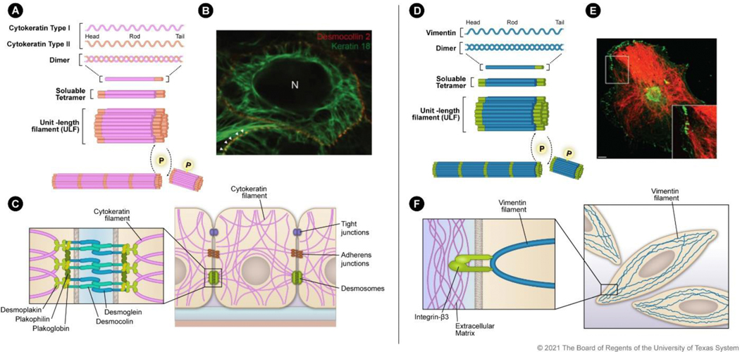
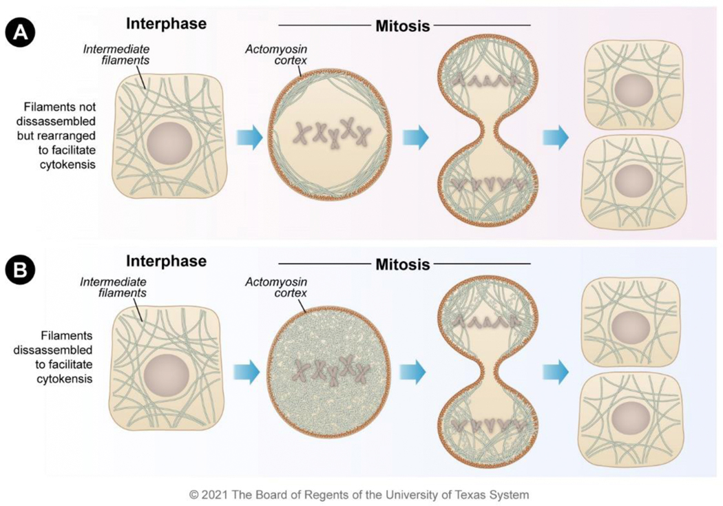
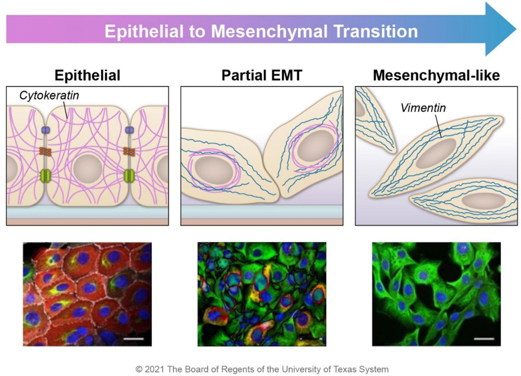
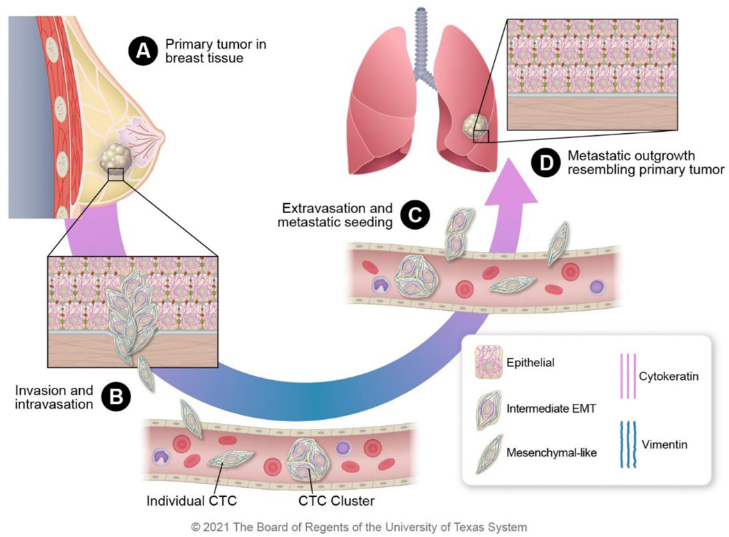
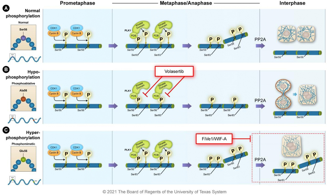
The cytoskeleton plays an integral role in maintaining the integrity of epithelial cells. Epithelial cells primarily employ cytokeratin in their cytoskeleton, whereas mesenchymal cells use vimentin. During the epithelial-mesenchymal transition (EMT), cytokeratin-positive epithelial cells begin to express vimentin. EMT induces stem cell properties and drives metastasis, chemoresistance, and tumor relapse. Most studies of the functions of cytokeratin and vimentin have relied on the use of either epithelial or mesenchymal cell types. However, it is important to understand how these two cytoskeleton intermediate filaments function when co-expressed in cells undergoing EMT. Here, we discuss the individual and shared functions of cytokeratin and vimentin that coalesce during EMT and how alterations in intermediate filament expression influence carcinoma progression.

Keywords: Cancer stem cells; Cytokeratin; Epithelial-mesenchymal transition; Intermediate filaments; Vimentin.


Conflict of interest statement

Declaration of Competing Interest The authors declare that they have no conflict of interest.

Figures

Figure 1.
Figure 1.. Filamentous assembly and localization of cytokeratin and vimentin:
A) Cytokeratin assembly initiates with type I and type II heterodimer formation. The heterodimers combine to form soluble tetramers that interact to form ULFs made up of eight monomers. ULFs are then assembled in a phosphorylation-dependent manner into 10-nm filaments. B) Survey fluorescence micrograph of MDCK cells that express YFP-labeled desmocollin (red) and mRFP-labeled human cytokeratin 18 (green). Image reprinted with permission from Quinlan et al., J. Cell Sci., 130, 3437–3445 (2017). C) Schematic of cytokeratin distribution in an epithelial cell with cell-cell adhesion processes. Zoomed inset shows the protein-protein interactions between cytokeratin filaments and desmosomes. D) Vimentin assembles as homodimers that combine to form soluble tetramers and then ULFs made up of eight monomers. ULFs are then assembled in a phosphorylation-dependent manner into filaments. E) Transformed human bone marrow endothelial cells stained for integrin β3 (green) and vimentin (red). Image reprinted with permission from Bhattacharya et al., J. Cell Sci., 122, 1390–1400 (2009). F) Schematic of vimentin distribution in a mesenchymal cell. Zoomed inset indicates the vimentin filament interaction with β3 integrin that anchors the filament to the extracellular matrix.
Figure 2.
Figure 2.. Localization mechanisms for vimentin and cytokeratin during mitosis.
Both vimentin and cytokeratin filaments exist as filamentous forms during interphase. During mitosis, the filaments either A) remain filamentous but are organized to the actin cortex to clear the mitotic plate or B) become disassembled into unit-length filaments to clear the mitotic plate. Both mechanisms enable the efficient completion of cytokinesis resulting in daughter cells.
Figure 3.
Figure 3.. Mutations in intermediate filament proteins result in mitotic defects in cell division.
A) Preventing phosphorylation through alanine mutations results in intermediate filaments that are unable to be disassembled during mitosis and cytokinesis. These filaments form an intermediate filament (IF) bridge between the daughter cells that can result in polyploidy. B) Phosphomimetic mutations or hyperphosphorylation of the intermediate filaments alters mitotic dynamics through tubulin localization and also leads to polyploidy.
Figure 4.
Figure 4.. Intermediate filament expression changes during EMT.
Epithelial-like cancer cells express cytokeratin (red), which aids in stress resistance and supports cell-cell adhesion; these cells do not express vimentin. As cells progress through EMT, mesenchymal markers such as vimentin (green) begin to be expressed. In E/M hybrid cells, cytokeratin is perinuclearly localized, and the vimentin filament network stretches from the nucleus to the periphery of the cell. At the far end of the EMT spectrum, epithelial markers are lost. The cartoons were constructed based on the observed morphology of HMLER cells during the transition from epithelial (CD104+ CD44low) to hybrid E/M (CD104+ CD44hi) to mesenchymal (CD104 CD44hi) populations . Images are reprinted with permission from Kröger, C. et al. Acquisition of a hybrid E/M state is essential for tumorigenicity of basal breast cancer cells. Proc. Natl. Acad. Sci. 116, 7353–7362 (2019).
Figure 5:
Figure 5:. Intermediate filament expression changes during cancer progression.
A) During primary tumor growth in the breast, some cells begin to undergo EMT. B) The cells with mesenchymal-like phenotypes function as leader cells during invasion and intravasation into the bloodstream. Individual circulating tumor cells (CTCs) and clusters of CTCs have both been observed in circulation. C) CTCs extravagate to metastatic sites to seed the tissues and remain dormant (in lungs in this example). D) At the metastatic site, the dormant carcinoma cells resume proliferation and ultimately resemble the primary tumor with largely epithelial morphology as a result of the mesenchymal to epithelial transition.
Figure 6.
Figure 6.. Small-molecule inhibitors have been identified that disrupt vimentin phospho-malleability.
A) During prometaphase, vimentin is phosphorylated at Ser56 by CDK1-cyclin B; this primes subsequent phosphorylation events. The polo-box domain (PDB) of PLK1 binds to the primed phosphorylation site at Ser56 and the kinase domain of PLK1 phosphorylates vimentin Ser83. This leads to the disassembly of the 10-nm filaments into ULFs during metaphase and anaphase. The phosphatase PP2A dephosphorylates vimentin stabilizing the filaments after cytokinesis. B) The PLK1 inhibitor volasertib binds to the ATP pocket of PLK1 preventing phosphorylation of substrates like vimentin. In the presence of volasertib, vimentin remains filamentous during metaphase causing issues during mitosis and cytokinesis that culminate in intermediate filament bridges and polyploidy. C) The small molecules FiVe1 and Withaferin-A (WIF-A) stabilize vimentin phosphorylation causing improper destabilization of vimentin filaments resulting in mitotic catastrophe and polyploidy.

References

    1. Snider NT & Omary MB Post-translational modifications of intermediate filament proteins: Mechanisms and functions. Nat. Rev. Mol. Cell Biol 15, 163–177 (2014). - PMC - PubMed
    1. Ivaska J. et al. PKCε-mediated phosphorylation of vimentin controls integrin recycling and motility. EMBO J. 24, 3834–3845 (2005). - PMC - PubMed
    1. Nishimura Y, Kasahara K. & Inagaki M. Intermediate filaments and IF-associated proteins: from cell architecture to cell proliferation. Proc. Jpn. Acad. Ser. B. Phys. Biol. Sci 95, 479–493 (2019). - PMC - PubMed
    1. Guo M. et al. The role of vimentin intermediate filaments in cortical and cytoplasmic mechanics. Biophys. J 105, 1562–1568 (2013). - PMC - PubMed
    1. Szeverenyi I. et al. The human intermediate filament database: Comprehensive information on a gene family involved in many human diseases. Hum. Mutat 29, 351–360 (2008). - PubMed

Publication types