. 2022 Apr;76(4):921-933.
doi: 10.1016/j.jhep.2021.12.014. Epub 2021 Dec 23.

Long non-coding RNA ACTA2-AS1 promotes ductular reaction by interacting with the p300/ELK1 complex

Affiliations

Long non-coding RNA ACTA2-AS1 promotes ductular reaction by interacting with the p300/ELK1 complex

Amaia Navarro-Corcuera et al. J Hepatol. 2022 Apr.

Abstract

Background & aims: Biliary disease is associated with a proliferative/fibrogenic ductular reaction (DR). p300 is an epigenetic regulator that acetylates lysine 27 on histone 3 (H3K27ac) and is activated during fibrosis. Long non-coding RNAs (lncRNAs) are aberrantly expressed in cholangiopathies, but little is known about how they recruit epigenetic complexes and regulate DR. We investigated epigenetic complexes, including transcription factors (TFs) and lncRNAs, contributing to p300-mediated transcription during fibrosis.

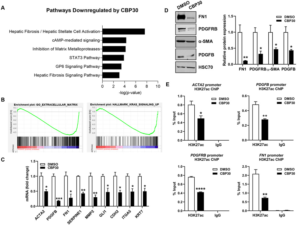
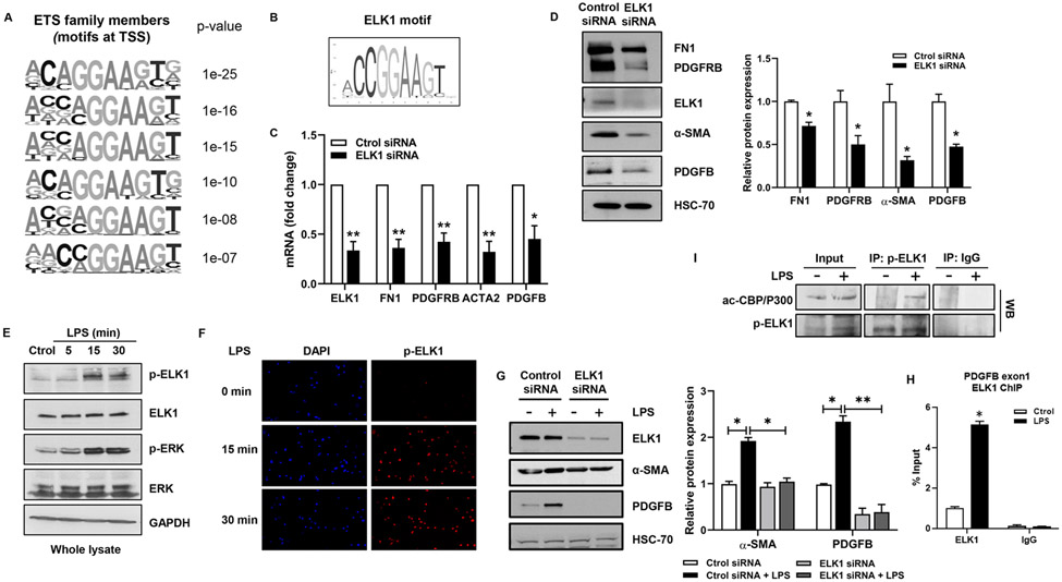
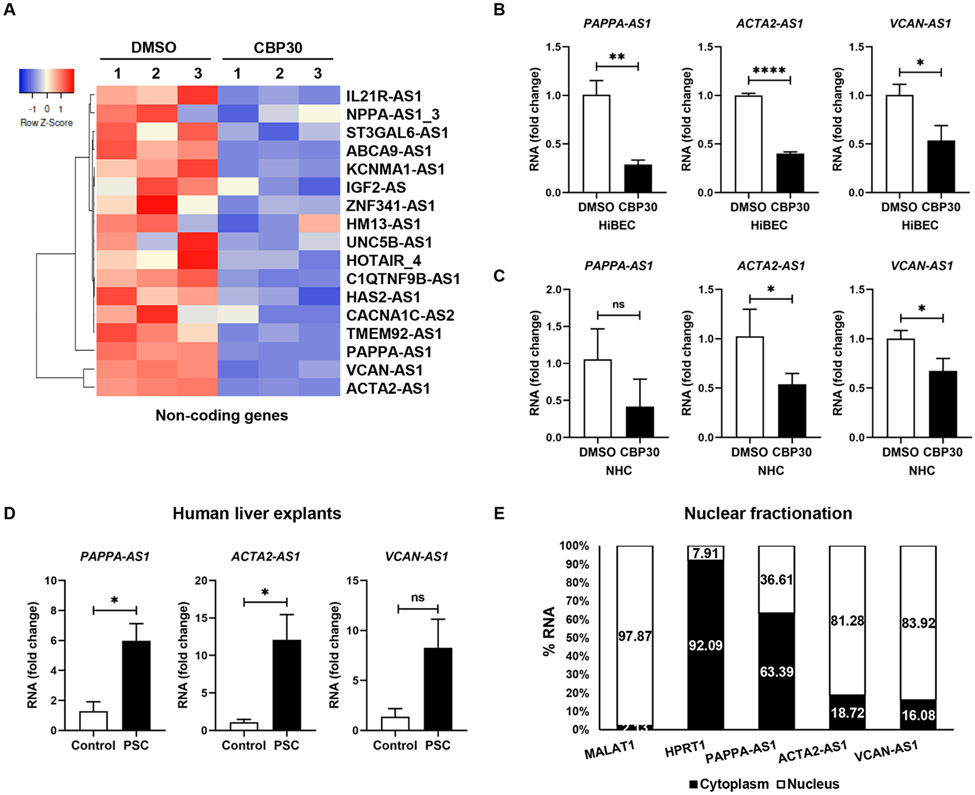
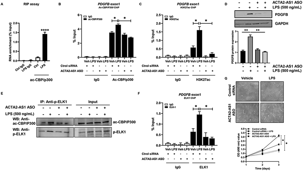
Methods: We evaluated p300 in vivo using tamoxifen-inducible, cholangiocyte-selective, p300 knockout (KO) coupled with bile duct ligation (BDL) and Mdr KO mice treated with SGC-CBP30. Primary cholangiocytes and liver tissue were analyzed for expression of Acta2-as1 lncRNA by qPCR and RNA in situ hybridization. In vitro, we performed RNA-sequencing in human cholangiocytes with a p300 inhibitor. Cholangiocytes were exposed to lipopolysaccharide (LPS) as an injury model. We confirmed formation of a p300/ELK1 complex by immunoprecipitation (IP). RNA IP was used to examine interactions between ACTA2-AS1 and p300. Chromatin IP assays were used to evaluate p300/ELK1 occupancy and p300-mediated H3K27ac. Organoids were generated from ACTA2-AS1-depleted cholangiocytes.

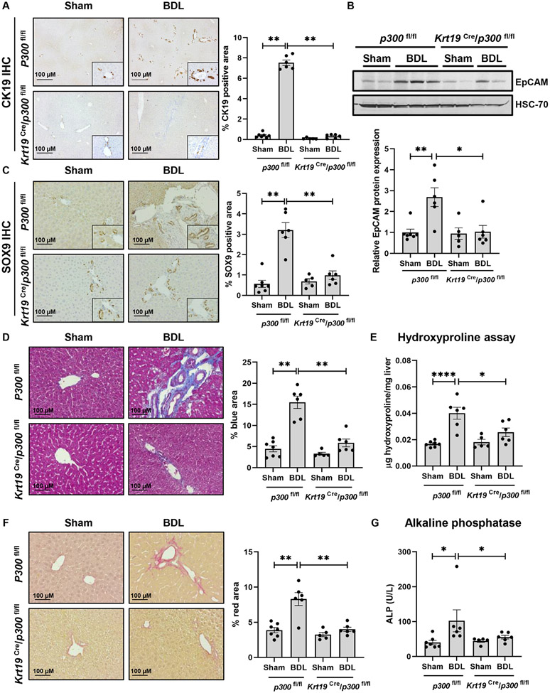
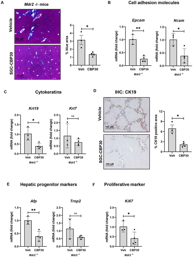
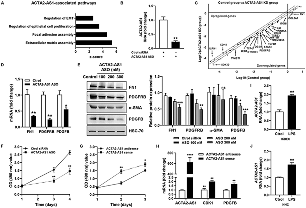
Results: BDL-induced DR and fibrosis were reduced in Krt19-CreERT/p300fl/fl mice. Similarly, Mdr KO mice were protected from DR and fibrosis after SGC-CBP30 treatment. In vitro, depletion of ACTA2-AS1 reduced expression of proliferative/fibrogenic markers, reduced LPS-induced cholangiocyte proliferation, and impaired organoid formation. ACTA2-AS1 regulated transcription by facilitating p300/ELK1 binding to the PDGFB promoter after LPS exposure. Correspondingly, LPS-induced H3K27ac was mediated by p300/ELK1 and was reduced in ACTA2-AS1-depleted cholangiocytes.

Conclusion: Cholangiocyte-selective p300 KO or p300 inhibition attenuate DR/fibrosis in mice. ACTA2-AS1 influences recruitment of p300/ELK1 to specific promoters to drive H3K27ac and epigenetic activation of proliferative/fibrogenic genes. This suggests that cooperation between epigenetic co-activators and lncRNAs facilitates DR/fibrosis in biliary diseases.

Lay summary: We identified a three-part complex containing an RNA molecule, a transcription factor, and an epigenetic enzyme. The complex is active in injured bile duct cells and contributes to activation of genes involved in proliferation and fibrosis.

Keywords: ACTA2-AS1; ELK1; cholangiocyte; ductular reaction; p300.


Conflict of interest statement

Conflict of interest The authors declare no conflicts of interest relevant to this work. Please refer to the accompanying ICMJE disclosure forms for further details.

Figures

Figure 1:
Figure 1:
Cholangiocyte- selective knockout of p300 attenuates DR and biliary fibrosis. (A) Immunohistochemistry for CK19; (B) Immunoblotting for EpCAM; (C) Immunohistochemistry for SOX9; (D) Masson’s Trichrome staining; (E) Hydroxyproline assays; (F) Picro-Sirius red staining; and (G) Serum ALP in p300fl/fl and Krt19Cre/p300fl/fl animals with sham or BDL surgery. (n=5- 7 animals/group). Graph bars represent SEM. (A-D),(F-G) Mann-Whitney test *p<0.05,**p<0.01;(E) one-way ANOVA with Bonferroni comparison *p<0.05,****p<0.001.
Figure 2:
Figure 2:
Inhibition of p300 attenuates DR and biliary fibrosis in Mdr−/− mice. (A) Masson’s Trichrome staining (Unpaired Student's t-test *p<0.05); (B-C) qRT-PCR for Epcam, Ncam, Krt19 and Krt7 (Unpaired Student's t-test *p<0.05,**p<0.01); (D) Immunohistochemistry for CK19 Mann-Whitney test *p<0.05); (E-F) qRT-PCR analysis for Afp, Trop2, and Ki-67 in liver from Mdr−/− mice treated with vehicle or SGC-CBP30 (Unpaired Student's t-test *p<0.05,**p<0.01). (n=3-4 animals/group). Graph bars represent SEM.
Figure 3:
Figure 3:
P300 activates transcription related to proliferation and fibrosis in cholangiocytes. (A) Pathways identified by IPA. (B) GSEA comparing control (DMSO) and P300-inhibited (CBP30) cells. (C) Validation by qRT- PCR. (D) Validation by WB. (E) ChIP-qPCR analysis in HiBEC treated with CBP30 or DMSO for H3K27ac at ACTA2, PDGFB, PDGFRB and FN1 promoters. (n=3). Graph bars represent SEM. Unpaired Student's t-test *p<0.05,**p<0.01,***p<0.005,****p<0.001 compared with DMSO group.
Figure 4:
Figure 4:
ELK1 regulates proliferative and fibrogenic genes in cholangiocytes. (A) ETS motifs identified by HOMER. (B) ELK1 motif from Jaspar. (C) qRT- PCR for ELK1, FN1, PDGFRB, ACTA2, and PDGFB in NHC transfected with control or ELK1 siRNA (Unpaired Student's t-test *p<0.05,**p<0.01 compared with control siRNA group). (D) WB for ELK1, FN1, PDGFRB, α-SMA and PDGFB in NHC transfected with control or ELK1 siRNA (Unpaired Student's t-test *p<0.05 compared with control siRNA group). (E) WB for p-ELK1, ELK1, p-ERK and ERK in NHC treated with LPS. (F) IF for p-ELK1 in NHC treated with LPS. (G) WB for ELK1, α-SMA and PDGFB in NHC transfected with control or ELK1 siRNA +/− LPS (one-way ANOVA with Bonferroni comparison *p<0.05,**p<0.01). (H) ChIP-qPCR in NHC treated with LPS. (I) WB on p-ELK1 and IgG immunoprecipitates from NHC +/− LPS (Unpaired Student’s t-test *p<0.05 compared with control group).(n=3). Graph bars represent SEM.
Figure 5:
Figure 5:
ACTA2-AS1 is regulated by p300 and overexpressed in PSC. (A) Heatmap shows expression of lncRNAs in HiBEC treated with SGC-CBP30 or DMSO. (B, C) Validation by qRT-PCR. (D) qRT-PCR for PAPPA-AS1, ACTA2-AS1, and VCAN-AS1 in liver from healthy and PSC patients. (E) Percentage of RNA in the nuclear and cytoplasmic fractions by qRT-PCR. MALAT1 lncRNA and HPRT1 as nuclear and cytoplasmic controls.(n=3). Graph bars represent SEM. Unpaired Student's t-test *p<0.05,**p<0.01,****p<0.001 compared with control group.
Figure 6:
Figure 6:
ACTA2-AS1 regulates proliferative and fibrogenic genes in cholangiocytes. (A) lncHUB shows pathways associated with ACTA2-AS1. (B) Analysis of ACTA2-AS1 expression in HiBEC transfected with control or ACTA2-AS1 antisense oligonucleotide (ASO). (C) Scatterplot showing gene changes in HiBEC transfected with control or ACTA2-AS1 ASO. (D) Validation by qPCR. (E) WB for FN1, PDGFRB, α-SMA and PDGFB. (F) Proliferation of control and ACTA2-AS1 knocked-down cells. (G) Proliferation of cells transfected with ACTA2-AS1 antisense and cells transfected with ACTA2-AS1 sense. (H) qRT- PCR for CDK1 and PDGFB in cells transfected with ACTA2-AS1 sense or antisense. (I, J) qRT- PCR for ACTA2-AS1 in HiBEC and NHC +/− LPS. (n=3). Graph bars represent SEM. Unpaired Student's t-test *p<0.05,**p<0.01,***p<0.005,****p<0.001 compared with control group.
Figure 7:
Figure 7:
ACTA2-AS1 mediates P300- and ELK1-dependent transcription. (A) RIP of ac-CBP/P300-bound ACTA2-AS1 in NHC +/−LPS (Unpaired Student's t-test ****p<0.001 compared with control group). (B, C) NHC transfected with control or ACTA2-AS1 ASO +/− LPS analyzed by ChIP for ac-CBP/P300 and H3K27ac on the PDGFB promoter. (D) WB for PDGFB in cells transfected with ACTA2-AS1 ASO following LPS. (E) WB on ac-CBP/P300, p-ELK1 and IgG immunoprecipitates from HiBEC transfected with ACTA2-AS1 ASO +/− LPS. (F) Cholangiocytes transfected with control or ACTA2-AS1 ASO +/− LPS analyzed by ChIP for ELK1 on the PDGFB promoter. (G) Proliferation of cells transfected with ACTA2-AS1 ASO +/− LPS. (B-D),(F-G) one-way ANOVA with Bonferroni comparison *p<0.05,**p<0.01.(n=3). Graph bars represent SEM.

References

    1. Banales JM, Huebert RC, Karlsen T, Strazzabosco M, LaRusso NF, Gores GJ. Cholangiocyte pathobiology. Nat Rev Gastroenterol Hepatol 2019;16:269–281. - PMC - PubMed
    1. Sato K, Marzioni M, Meng F, Francis H, Glaser S, Alpini G. Ductular Reaction in Liver Diseases: Pathological Mechanisms and Translational Significances. Hepatology 2019;69:420–430. - PMC - PubMed
    1. Ko S, Russell JO, Molina LM, Monga SP. Liver Progenitors and Adult Cell Plasticity in Hepatic Injury and Repair: Knowns and Unknowns. Annual review of pathology 2020;15:23–50. - PMC - PubMed
    1. Lazaridis KN, LaRusso NF. Primary Sclerosing Cholangitis. N Engl J Med 2016;375:1161–70. - PMC - PubMed
    1. Jalan-Sakrikar N, De Assuncao TM, Shi G, Aseem SO, Chi C, Shah VH, et al. Proteasomal Degradation of Enhancer of Zeste Homologue 2 in Cholangiocytes Promotes Biliary Fibrosis. Hepatology 2019;70:1674–1689. - PMC - PubMed

Publication types