. 2021 Dec;48(12):1111-1121.
doi: 10.1016/j.jgg.2021.12.003. Epub 2021 Dec 24.

Evidence for a mouse origin of the SARS-CoV-2 Omicron variant

Affiliations

Evidence for a mouse origin of the SARS-CoV-2 Omicron variant

Changshuo Wei et al. J Genet Genomics. 2021 Dec.

Abstract

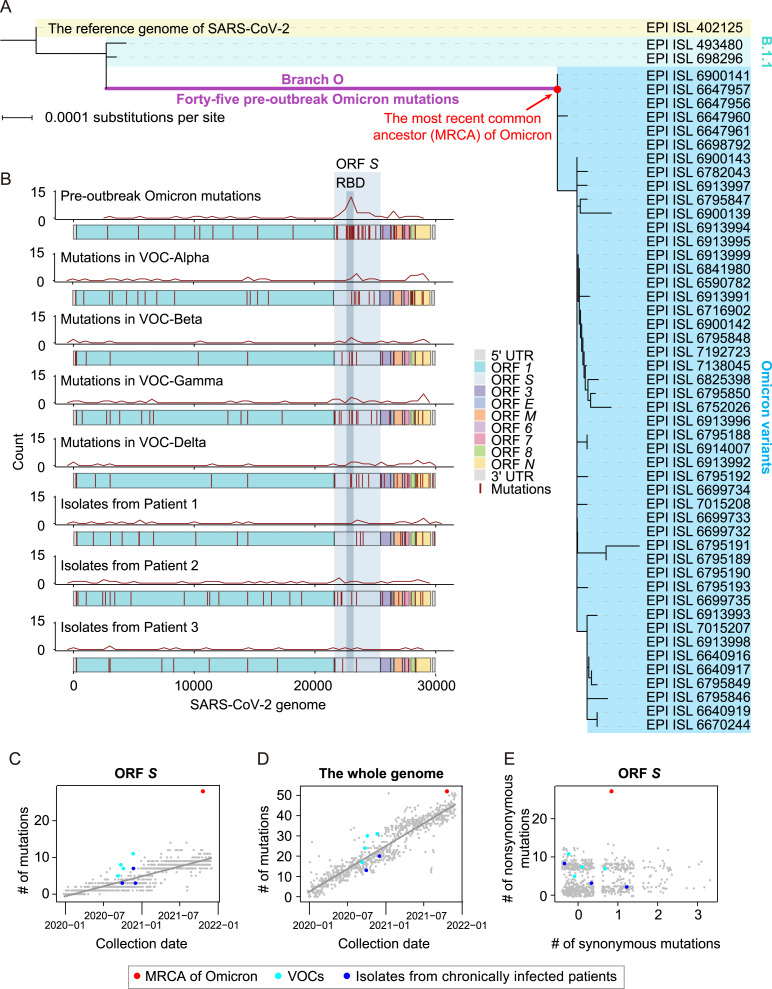
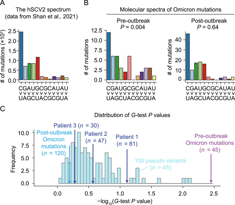
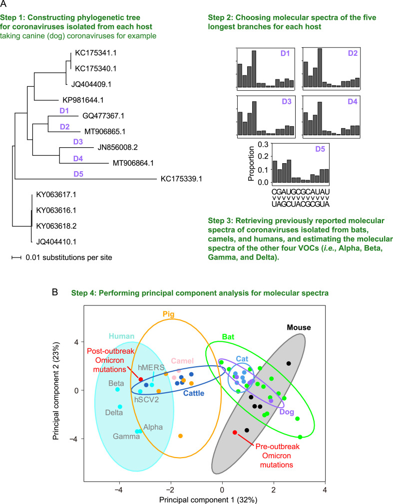
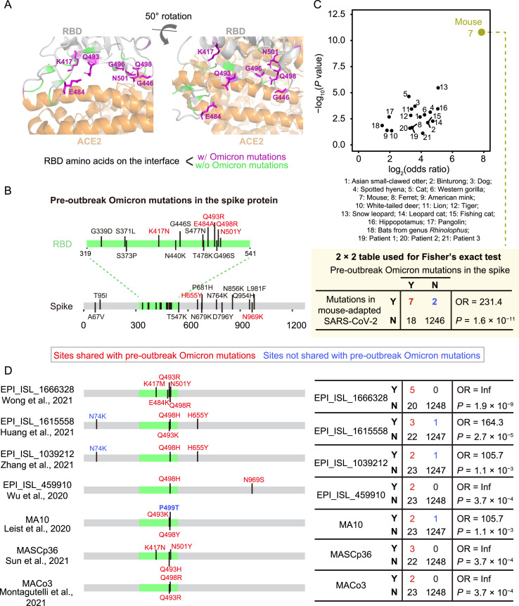
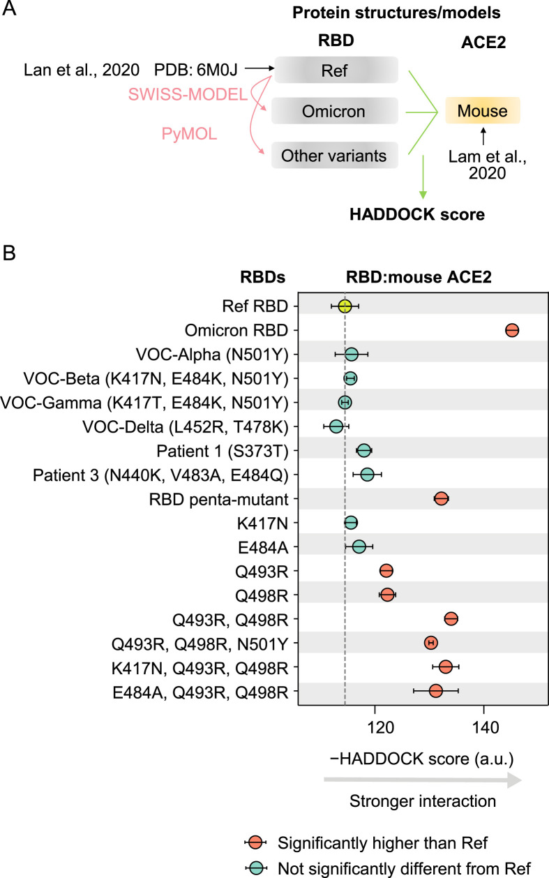
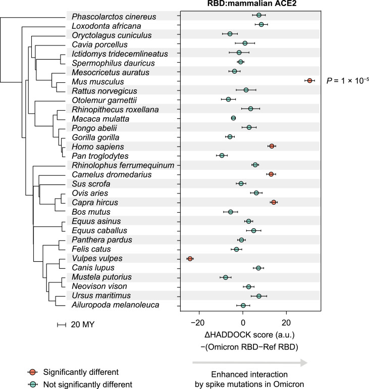
The rapid accumulation of mutations in the SARS-CoV-2 Omicron variant that enabled its outbreak raises questions as to whether its proximal origin occurred in humans or another mammalian host. Here, we identified 45 point mutations that Omicron acquired since divergence from the B.1.1 lineage. We found that the Omicron spike protein sequence was subjected to stronger positive selection than that of any reported SARS-CoV-2 variants known to evolve persistently in human hosts, suggesting a possibility of host-jumping. The molecular spectrum of mutations (i.e., the relative frequency of the 12 types of base substitutions) acquired by the progenitor of Omicron was significantly different from the spectrum for viruses that evolved in human patients but resembled the spectra associated with virus evolution in a mouse cellular environment. Furthermore, mutations in the Omicron spike protein significantly overlapped with SARS-CoV-2 mutations known to promote adaptation to mouse hosts, particularly through enhanced spike protein binding affinity for the mouse cell entry receptor. Collectively, our results suggest that the progenitor of Omicron jumped from humans to mice, rapidly accumulated mutations conducive to infecting that host, then jumped back into humans, indicating an inter-species evolutionary trajectory for the Omicron outbreak.

Keywords: Evolutionary origins; Molecular spectrum of mutations; Omicron; Receptor-binding domain; SARS-CoV-2; Spike-ACE2 interaction.


Conflict of interest statement

Conflict of interest The authors declare that they have no competing interests.

Figures

Fig. 1
Fig. 1
The characterization of pre-outbreak Omicron mutations. A: The phylogenetic tree of Omicron variants, including the reference genome of SARS-CoV-2, two B.1.1 variants, and 48 Omicron variants. A total of 45 pre-outbreak Omicron point mutations in the long branch (Branch O, labeled in purple) leading to the MRCA of Omicron (dot in red) in the phylogenetic tree are shown in Fig. S1. B: The genomic distribution of the 45 pre-outbreak Omicron mutations, the mutations detected in each progenitor of the other four VOCs (i.e., Alpha, Beta, Gamma, and Delta), and the mutations identified from the SARS-CoV-2 isolates of three chronically infected patients. The density curves for mutations were generated by the geom_freqpoly function in R. C: Number of mutations that accumulated in ORF S of the MRCA of Omicron (red), the other four VOCs (cyan), and three SARS-CoV-2 isolates from chronically infected patients (blue), against the date of sample collection. SARS-CoV-2 variants randomly sampled (one variant per day) are shown in grey, and the grey line represents their linear regression. D: Similar to (C), for the whole genome. The dot for a variant isolated from a chronically infected patient is overlapped with a VOC. E: A scatterplot shows the numbers of synonymous and nonsynonymous mutations in ORF S (jittered in order to reduce overplotting). UTR, untranslated region; RBD, receptor-binding domain.
Fig. 2
Fig. 2
Comparison of the molecular spectrum of pre-outbreak Omicron mutations and spectra of mutations known to accumulate in humans. A: The molecular spectrum of viral mutations that accumulated in humans (the hSCV2 spectrum). B: The molecular spectra of pre-outbreak and post-outbreak Omicron mutations. P values were given by G-tests against the hSCV2 spectrum. C: The distribution of P values (given by G-tests) of 100 pseudo samples that were downsampled from the hSCV2 spectrum. The number of mutations (n) of each pseudo sample was equal to 45. SARS-CoV-2 data of Patient 1 were retrieved from Kemp et al. (2021), and those of Patients 2 and 3 were retrieved from Truong et al. (2021).
Fig. 3
Fig. 3
The similarity in molecular spectra between Omicron and coronaviruses isolated from various mammalian species. A: A schematic shows the workflow for analyzing the similarity in molecular spectra across various hosts. B: The principal component analysis plot depicts the molecular spectra of virus mutations that accumulated in humans and various host species. Dots were colored according to the corresponding host species. The 95% confidence ellipses are shown for each host species. VOC, variant of concern.
Fig. 4
Fig. 4
The similarity in the spike protein sequence between Omicron and SARS-CoV-2 variants isolated from various hosts. A: The interface structure between SARS-CoV-2 spike protein and human ACE2 (PDB: 6M0J). RBD residues on the interface (with a distance cut-off of 5 Å) were labeled. B: The pre-outbreak Omicron mutations in the spike protein. C: The statistical assessment on the overlapping in mutated positions of the spike protein between Omicron and SARS-CoV-2 variants isolated from chronically infected patients or nonhuman mammals using Fisher’s exact tests. The 2 × 2 contingency table for mice is shown. D: Comparison between pre-outbreak Omicron mutations and mutations detected in seven mouse-adapted SARS-CoV-2 variants in the spike protein. OR, odds ratio; Y, with a mutation at a particular site; N, without a mutation at the site.
Fig. 5
Fig. 5
Predicted binding affinities between RBD variants and mouse ACE2. A: A schematic shows the workflow to estimate the HADDOCK scores between RBD variants and mouse ACE2. B: The HADDOCK scores for the interaction of various RBD variants with the mouse ACE2. The error bars represent standard errors. Penta-mutant of RBD harbored five mutations (K417N, E484A, Q493R, Q498R, and N501Y). The result of Patient 2, who did not harbor any amino acid mutations in RBD, was not shown.
Fig. 6
Fig. 6
Predicted interaction enhancement with ACE2 of various mammalian species caused by pre-outbreak Omicron mutations in the spike protein. P values were given by two-tailed t-tests. The phylogenetic tree was constructed using TimeTree, in the unit of MY. MY, million years.

References

    1. Ashkenazy H., Penn O., Doron-Faigenboim A., Cohen O., Cannarozzi G., Zomer O., Pupko T. FastML: a web server for probabilistic reconstruction of ancestral sequences. Nucleic Acids Res. 2012;40:W580–W584. - PMC - PubMed
    1. Blanc V., Davidson N.O. APOBEC-1-mediated RNA editing. Wiley Interdiscip. Rev. Syst. Biol. Med. 2010;2:594–602. - PMC - PubMed
    1. Callaway E. Heavily mutated Omicron variant puts scientists on alert. Nature. 2021 doi: 10.1038/d41586-021-03552-w. - DOI - PubMed
    1. Cameroni E., Bowen J.E., Rosen L.E., Saliba C., Zepeda S.K., Culap K., Pinto D., VanBlargan L.A., Marco A.D., Di Iulio J., et al. Broadly neutralizing antibodies overcome SARS-CoV-2 Omicron antigenic shift. Nature. 2021 doi: 10.1038/d41586-021-03825-4. - DOI - PMC - PubMed
    1. Chandler J.C., Bevins S.N., Ellis J.W., Linder T.J., Tell R.M., Jenkins-Moore M., Root J.J., Lenoch J.B., Robbe-Austerman S., DeLiberto T.J., et al. SARS-CoV-2 exposure in wild white-tailed deer (Odocoileus virginianus) Proc. Natl. Acad. Sci. U. S. A. 2021;118 - PMC - PubMed

Substances

Supplementary concepts