Review
. 2022 Jan:82:105887.
doi: 10.1016/j.ultsonch.2021.105887. Epub 2021 Dec 22.

Sonoproduction of nanobiomaterials - A critical review

Affiliations
Review

Sonoproduction of nanobiomaterials - A critical review

Sze Shin Low et al. Ultrason Sonochem. 2022 Jan.

Abstract

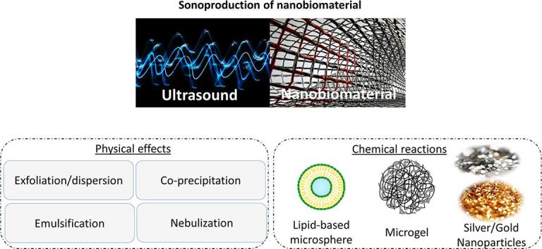
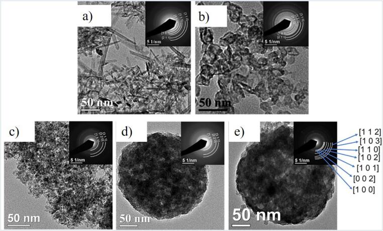
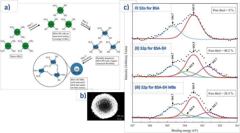
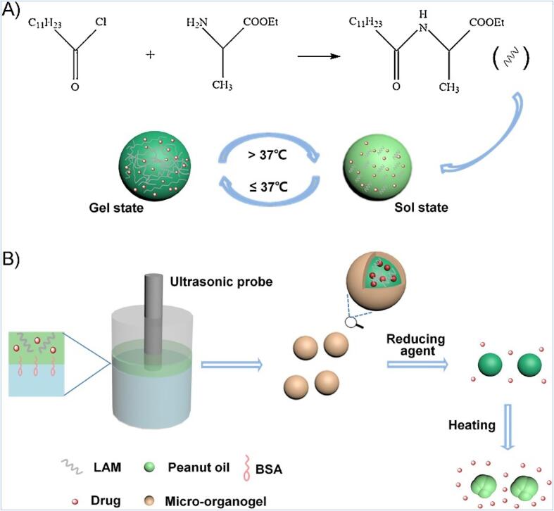
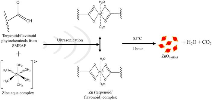
Ultrasound (US) demonstrates remarkable potential in synthesising nanomaterials, particularly nanobiomaterials targeted towards biomedical applications. This review briefly introduces existing top-down and bottom-up approaches for nanomaterials synthesis and their corresponding synthesis mechanisms, followed by the expounding of US-driven nanomaterials synthesis. Subsequently, the pros and cons of sono-nanotechnology and its advances in the synthesis of nanobiomaterials are drawn based on recent works. US-synthesised nanobiomaterials have improved properties and performance over conventional synthesis methods and most essentially eliminate the need for harsh and expensive chemicals. The sonoproduction of different classes and types of nanobiomaterials such as metal and superparamagnetic nanoparticles (NPs), lipid- and carbohydrate-based NPs, protein microspheres, microgels and other nanocomposites are broadly categorised based on the physical and/or chemical effects induced by US. This review ends on a good note and recognises US-driven synthesis as a pragmatic solution to satisfy the growing demand for nanobiomaterials, nonetheless some technical challenges are highlighted.

Keywords: Cavitation; Material; Nano; Nanobiomaterial; Sonoproduction; Synthesis; Ultrasound.


Conflict of interest statement

The authors declare that they have no known competing financial interests or personal relationships that could have appeared to influence the work reported in this paper.

Figures

None
Graphical abstract
Fig. 1
Fig. 1
(a) Simulated changes in temperature and radius of an SBSL bubble at steady state, where the collapse occurred within 0.1 ms (b) Calculated results of the number of molecules inside a bubble based on the SBSL model. Various species are formed as high heat from the cavitation dissociates air and water vapour within the bubble. Reprinted from with permission from AIP Publishing.
Fig. 2
Fig. 2
(a, b) SEM images of exfoliated graphene. (c) TEM image of Herceptin-graphene (d) Schematic image of the synthesised graphene. Reprinted from with permission from the American Chemical Society.
Fig. 3
Fig. 3
Formation illustration of Fe3O4 nanocomposites via (a) shaking assisted process and (b) US irradiation. TEM images of (c,d) multi-core nanocomposites show the composite having a polycrystalline core while (e,f) nanocomposites produced from US assistance exhibit clear mono core–shell structure, attributed to the diffusive and surface-protective shockwaves from implosive bubble collapse that form discrete core Fe3O4 NPs within silica. (g) In the US-assisted preparation of MNPs, it has been hypothesised that the shock waves generated from the collapse of the acoustic bubble counteract the high tension of MNPs, thus preventing crystalline coalescence on neighbouring surfaces that would yield heterogeneous-sized products. Reprinted from with permission from Elsevier (CC-BY license).
Fig. 4
Fig. 4
TEM images of (a) rod-like and (b) spherical ZnF NPs (c) pristine ZnS (d) sphalerite-rich N-doped ZnS and (e) wurtzite-rich N-doped ZnS NPs. Insets are Selected Area Electron Diffraction (SAED) patterns corresponding to XRD patterns. Reprinted from with permission from Elsevier.
Fig. 5
Fig. 5
(a) Schematic and the flow chart exhibiting the synthesis of surface-treated BSA MBs (BSA-SH-MBs) (b) SEM image of spherical BSA-SH-MBs (c) High-resolution XPS spectra of S2P for (ci) BSA protein before surface treatment (cii) BSA-SH post-treatment and (ciii) cross-linked MBs BSA-SH-MBs illustrate an increase in free thiol after treatment and decreased after cross-linking of the microsphere. Reprinted from with permission from Elsevier.
Fig. 6
Fig. 6
Schematic illustration of (A) sol–gel of gelator LAM through the reaction between acyl chloride and esterified L-amino acid, where the aggregates formed upon cooling (B) sonochemical assembly of drug- encapsulated BSA-MBs and stimuli-triggered release of the drug-loaded micro-organogel. Reprinted from with permission from Elsevier.
Fig. 7
Fig. 7
Proposed mechanism for the US-assisted bioreduction of zinc salt precursor into ZnO nanorice with phytochemicals from SMEAF. Reprinted from with permission from de Gruyter (CC-BY license).
Fig. 8
Fig. 8
Biological reduction of Zn ions under sonication yielded ZnO with nanorice structure. FESEM micrographs of (a-c) ZnOChem (d-f) ZnOSMEAF retained a similar structure with the addition of SMEAF; STEM micrographs of (g) ZnOChem (h) ZnOSMEAF of scale bar 200 and 500 nm, respectively. Reprinted from with permission from de Gruyter (CC-BY license).
Fig. 9
Fig. 9
FTIR spectra of (a) SMEAF (b) ZnOSMEAF and (c) ZnOChem. Phytochemicals in SMEAF, particularly OH– groups in flavonoids, behave as reducing and capping agents controlling the size and morphology of ZnO NPs as indicated in the wide peak at 365 and 3250 cm−1 from O-H stretching vibration. Reprinted from with permission from de Gruyter (CC-BY license).
Fig. 10
Fig. 10
Powder X-ray diffraction (PXRD) analyses of (a) Fe3O4 NPs (b) bio-MOF single crystal (c) bio-MOF nanostructures, indicated higher crystallinity of pre-synthesised NP and sonochemically produced bio-MOFs (d) magnetic bio-MOF nanocomposites retained similar diffraction peaks as prior to the addition of Fe3O4, showing no obvious effect on the bio-MOF framework. Reprinted from with permission from Elsevier.
Fig. 11
Fig. 11
FESEM images of sonochemically synthesised Fe3O4@bio-MOF nanostructures at various times of sonication (a) 30 min (b) 60 min (c) 90 min and (d) 120 min. Sonication of up to 120 min was deemed the optimal time parameter that produced a non-agglomerated porous-layer open morphology. In contrast, different sizes and agglomerated Fe3O4 particles were observed within the shorter period. Reprinted from with permission from Elsevier.

References

    1. Mishra R.K., Ha S.K., Verma K., Tiwari S.K. Recent progress in selected bio-nanomaterials and their engineering applications: an overview. J. Sci. Adv. Mater. Dev. 2018;3(3):263–288. doi: 10.1016/j.jsamd.2018.05.003. - DOI
    1. McNamara K., Tofail S.A.M. Nanosystems: the use of nanoalloys, metallic, bimetallic, and magnetic nanoparticles in biomedical applications. Phys. Chem. Chem. Phys. 2015;17(42):27981–27995. doi: 10.1039/C5CP00831J. - DOI - PubMed
    1. Oliveira O.N., Iost R.M., Siqueira J.R., Crespilho F.N., Caseli L. Nanomaterials for diagnosis: challenges and applications in smart devices based on molecular recognition. ACS Appl. Mater. Interfaces. 2014;6(17):14745–14766. doi: 10.1021/am5015056. - DOI - PubMed
    1. Patra J.K., Das G., Fraceto L.F., Campos E.V.R., Rodriguez-Torres M.D.P., Acosta-Torres L.S., Diaz-Torres L.A., Grillo R., Swamy M.K., Sharma S., Habtemariam S., Shin H.-S. Nano based drug delivery systems: recent developments and future prospects. J. Nanobiotechnol. 2018;16(1) doi: 10.1186/s12951-018-0392-8. - DOI - PMC - PubMed
    1. Torres-Sangiao E., Holban A.M., Gestal M.C. Advanced nanobiomaterials: vaccines, diagnosis and treatment of infectious diseases. Molecules. 2016;21:867. doi: 10.3390/molecules21070867. - DOI - PMC - PubMed

LinkOut - more resources