Review
. 2022 Mar 1;33(2):80-90.
doi: 10.1097/ICU.0000000000000829.

Glaucoma and biomechanics

Affiliations
Review

Glaucoma and biomechanics

Babak N Safa et al. Curr Opin Ophthalmol. .

Abstract

Purpose of review: Biomechanics is an important aspect of the complex family of diseases known as the glaucomas. Here, we review recent studies of biomechanics in glaucoma.

Recent findings: Several tissues have direct and/or indirect biomechanical roles in various forms of glaucoma, including the trabecular meshwork, cornea, peripapillary sclera, optic nerve head/sheath, and iris. Multiple mechanosensory mechanisms and signaling pathways continue to be identified in both the trabecular meshwork and optic nerve head. Further, the recent literature describes a variety of approaches for investigating the role of tissue biomechanics as a risk factor for glaucoma, including pathological stiffening of the trabecular meshwork, peripapillary scleral structural changes, and remodeling of the optic nerve head. Finally, there have been advances in incorporating biomechanical information in glaucoma prognoses, including corneal biomechanical parameters and iridial mechanical properties in angle-closure glaucoma.

Summary: Biomechanics remains an active aspect of glaucoma research, with activity in both basic science and clinical translation. However, the role of biomechanics in glaucoma remains incompletely understood. Therefore, further studies are indicated to identify novel therapeutic approaches that leverage biomechanics. Importantly, clinical translation of appropriate assays of tissue biomechanical properties in glaucoma is also needed.


Conflict of interest statement

Conflict of interest

Authors declare no conflict of interest.

Figures

Figure 1
Figure 1
Schematic of the human eye. The biomechanics of the eye is closely related to pathophysiology of glaucoma. In the anterior section of the globe, the outflow of aqueous humor through the trabecular meshwork (TM) and Schlemm’s canal (SC) is affected by the mechanical properties of the TM; for example, in primary open-angle glaucoma (POAG) the stiffness of TM is increased, likely due to biological remodeling of the TM extracellular matrix (ECM) under elevated IOP. Additionally, new devices that measure the biomechanics of the cornea may aid in monitoring the risk of glaucoma. Further, the biomechanics of the iris is a new topic of interest in primary angle-closure glaucoma (PACG), where the role of tissue mechanical properties and muscular contractions of the iris are important. In the posterior globe, the sclera’s biomechanics, especially the peripapillary sclera, is known to strongly influence the mechanical loads on the optic nerve head (ONH). Lastly, the mechanical properties of the ONH and lamina cribrosa (LC) and their pathological remodeling in POAG (i.e., cupping) are important aspects of RGC loss in glaucoma, and are associated with mechanobiological remodeling of the ONH by resident cells, especially astrocytes and LC cells. (Figure reproduced from Paula et al. 2016 [136] with permission from the publisher.)
Figure 2
Figure 2
Estimation of TM stiffness in mice with induced steroid glaucoma. (A-F) Spectral domain-optical coherence tomography (SD-OCT) images of the limbal region in living C57BL/6 mouse eyes at different imposed IOP levels. Eyes were treated with netarsudil, an FDA-approved rho-kinase inhibitor, or placebo. The blue shading shows the automatically identified SC lumen. (G) Estimation of TM stiffness based on OCT images by inverse finite element modeling (iFEM), showing a stiff TM in a steroid glaucoma model, with restoration of normal TM stiffness by netarsudil. (H) TM stiffness directly measured by atomic force microscopy (AFM) in mice receiving steroid and netarsudil or placebo. DEX = Dexamethasone, NT=netarsudil, PL=placebo (Figure reproduced from Li et al. 2021 [33].)
Figure 3
Figure 3
The use of the Ocular Response Analyzer (ORA) in glaucoma. (A) Schematic of typical data from the ORA, in which an air pulse is delivered to the cornea (green) and corneal deformation is measured by a photodetector (red). Corneal hysteresis (mmHg) is defined as the difference between the applanation pressure 1, as the cornea moves inwards, and applanation pressure 2, as it returns to its normal shape. (B) Cumulative probability of glaucoma development in two groups of OH subjects classified by corneal hysteresis (CH) values measured with an ORA. (A) is reproduced from Kaushik et al. [158], and (B) is reproduced from Susanna et al. [159] with permission from the publishers.
Figure 4
Figure 4
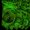
Optical imaging was used to identify the trajectories of collagen fibers in the ONH and peripapillary sclera of a post mortem human eye. One can observe both (+) radial and (∼) circumferential fiber orientations, as well as the less aligned interweaving regions of the sclera (right side of image). The fibrous lamina cribrosa, within the scleral canal, can also be seen on the left side of the image. (Reproduced from Gogola et al. [164].)
Figure 5
Figure 5
Schematic showing the major loads acting on the tissues of the optic nerve head. The IOP acts directly at the vitreoretinal interface to posteriorly displace ONH tissues (dark blue arrows), as well as enlarging the eye globe and thus causing tension in the scleral wall which is transmitted to the ONH (red arrows). The cerebrospinal fluid (CSF) is also pressurized (purple) which in turn causes a retrolaminar tissue (RLT) pressure that anteriorly displaces the lamina cribrosa (light blue arrows). Not shown is tension in the optic nerve sheath. Modified from Albon et al. [165], from a scanning electron micrograph of the connective tissue of a human ONH.
Figure 6
Figure 6
Iris deformation is shown under (A) normal and (B) dim light conditions using anterior segment OCT. Using inverse finite element modeling (C and D), where the stroma [blue region] is deformed by the sphincter smooth muscle [red region] Pant and co-workers calculated the mechanical properties of the iris, and showed that subjects with a history of PACG (post-LPI) tend to have a c. 3 times greater stromal stiffness compared to healthy subjects (Figure adapted from Pant et al. [152] with permission from the publisher.)

References

    1. Heijl A, Leske MC, Bengtsson B, et al., “Reduction of intraocular pressure and glaucoma progression: results from the Early Manifest Glaucoma Trial,” Arch Ophthalmol, vol. 120, no. 10, pp. 1268–1279, 2002, doi: ecs20122 [pii]. - PubMed
    1. Leske MC, Heijl A, Hussein M, et al., “Factors for glaucoma progression and the effect of treatment: the early manifest glaucoma trial,” Arch Ophthalmol, vol. 121, no. 1, pp. 48–56, 2003, doi: ecs20139 [pii]. - PubMed
    1. Anderson DR, Drance SM, and Schulzer M, “Natural history of normal-tension glaucoma,” Ophthalmology, vol. 108, no. 2, pp. 247–253, 2001, doi: S0161642000005182 [pii]. - PubMed
    1. Gaasterland DE, Ederer F, Beck A, et al., “The Advanced Glaucoma Intervention Study (AGIS): 7. The relationship between control of intraocular pressure and visual field deterioration,” Am. J. Ophthalmol, vol. 130, no. 4, pp. 429–440, 2000, doi: 10.1016/S0002-9394(00)00538-9. - DOI - PubMed
    1. Stowell C, Burgoyne CF, Tamm ER, et al., “Biomechanical aspects of axonal damage in glaucoma: A brief review,” Exp. Eye Res, vol. 157, pp. 13–19, Apr. 2017, doi: 10.1016/j.exer.2017.02.005. - DOI - PMC - PubMed