. 2022 Feb;91(2):225-237.
doi: 10.1002/ana.26293. Epub 2022 Jan 20.

Variants in Mitochondrial ATP Synthase Cause Variable Neurologic Phenotypes

Affiliations

Variants in Mitochondrial ATP Synthase Cause Variable Neurologic Phenotypes

Michael Zech et al. Ann Neurol. 2022 Feb.

Abstract

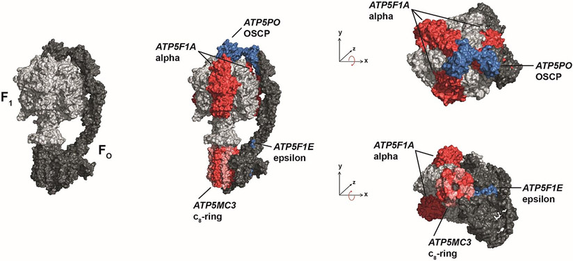
Objective: ATP synthase (ATPase) is responsible for the majority of ATP production. Nevertheless, disease phenotypes associated with mutations in ATPase subunits are extremely rare. We aimed at expanding the spectrum of ATPase-related diseases.

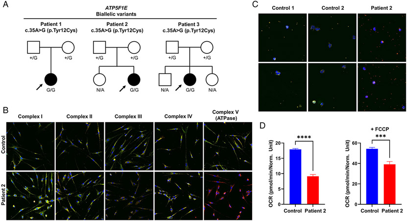
Methods: Whole-exome sequencing in cohorts with 2,962 patients diagnosed with mitochondrial disease and/or dystonia and international collaboration were used to identify deleterious variants in ATPase-encoding genes. Findings were complemented by transcriptional and proteomic profiling of patient fibroblasts. ATPase integrity and activity were assayed using cells and tissues from 5 patients.

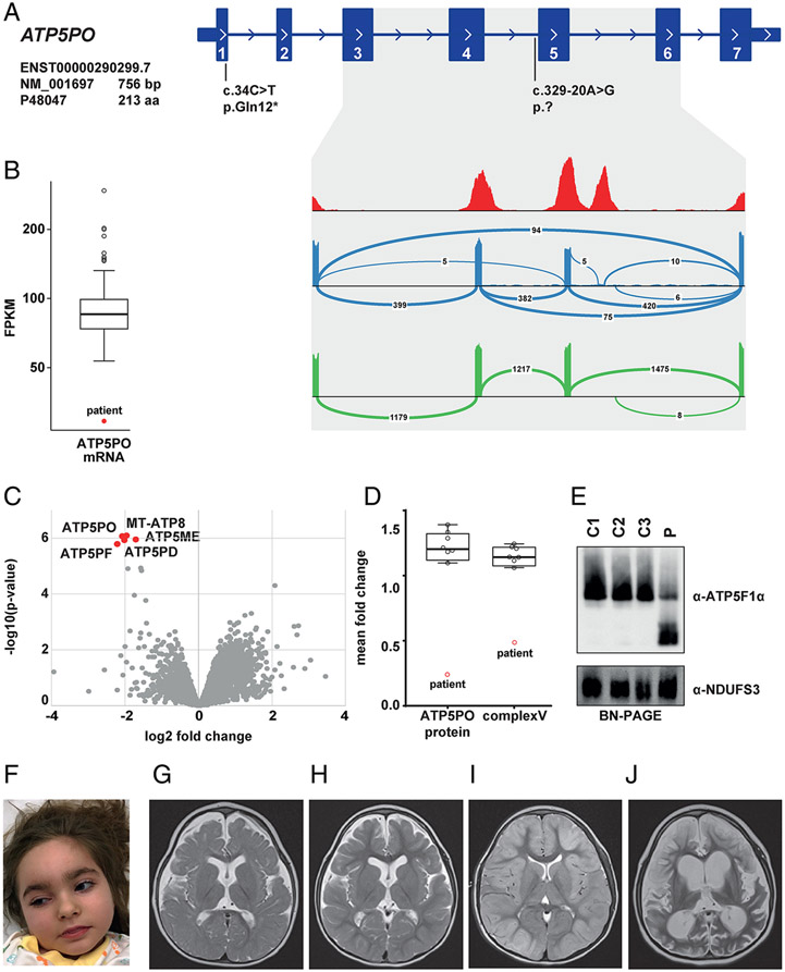
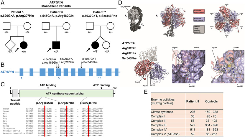
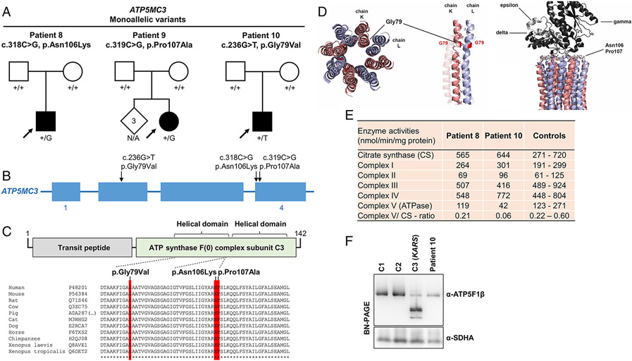
Results: We present 10 total individuals with biallelic or de novo monoallelic variants in nuclear ATPase subunit genes. Three unrelated patients showed the same homozygous missense ATP5F1E mutation (including one published case). An intronic splice-disrupting alteration in compound heterozygosity with a nonsense variant in ATP5PO was found in one patient. Three patients had de novo heterozygous missense variants in ATP5F1A, whereas another 3 were heterozygous for ATP5MC3 de novo missense changes. Bioinformatics methods and populational data supported the variants' pathogenicity. Immunohistochemistry, proteomics, and/or immunoblotting revealed significantly reduced ATPase amounts in association to ATP5F1E and ATP5PO mutations. Diminished activity and/or defective assembly of ATPase was demonstrated by enzymatic assays and/or immunoblotting in patient samples bearing ATP5F1A-p.Arg207His, ATP5MC3-p.Gly79Val, and ATP5MC3-p.Asn106Lys. The associated clinical profiles were heterogeneous, ranging from hypotonia with spontaneous resolution (1/10) to epilepsy with early death (1/10) or variable persistent abnormalities, including movement disorders, developmental delay, intellectual disability, hyperlactatemia, and other neurologic and systemic features. Although potentially reflecting an ascertainment bias, dystonia was common (7/10).

Interpretation: Our results establish evidence for a previously unrecognized role of ATPase nuclear-gene defects in phenotypes characterized by neurodevelopmental and neurodegenerative features. ANN NEUROL 2022;91:225-237.


Conflict of interest statement

Potential Conflicts of Interest

None of the authors has any relevant conflict of interest to declare.

Figures

Figure 1:
Figure 1:
Schematic views illustrating the subunits of mitochondrial ATPase mutated in patients of this study. ATPase consists of two functional domains, FO (dark gray) and F1 (light gray). Variant-bearing subunits are shown in red and blue colors: ε-subunit (encoded by ATP5F1E); OSCP-subunit (encoded by ATP5PO); α-subunit (encoded by ATP5F1A); and subunit c (encoded by ATP5MC3).
Figure 2:
Figure 2:. Biallelic ATP5F1E variants identified in 3 cases
(a) Pedigree drawings for 3 families with the homozygous variant in ATP5F1E. N/A, no genotyping available. (b) Fluorescence staining of patient 2`s primary human skin fibroblasts and a fibroblast control line. Cells were stained with antibodies raised against subunits of the five OXPHOS complexes (green) and an antibody against VDAC1 (red). The merge is shown. A shift to red indicates a reduction of the respective OXPHOS subunit. The images were taken with a 200x magnification. The experiment was performed with analysis of six different control lines (data not shown) and repeated twice with similar results. (c) Fluorescence staining of blood smears from patient 2 and two different control lines. Cells were stained with an antibody raised against the ATP5F1A subunit of ATPase (green) and an antibody against VDAC1 (red). The merge is shown. A shift to red indicates a reduction of the ATP5F1A subunit. The images were taken with a 400x magnification. The experiment was repeated twice with similar results. (d) Measurement of oxygen consumption rate (OCR) in patient 2`s fibroblasts and a control line. Data show the mean values ± SEM of basal respiration (left graph) and of maximal respiration after addition of FCCP (right graph); number of replicates: patient=4; control=8; ***p<0.001, ****p<0.0001.
Figure 3:
Figure 3:. Biallelic ATP5PO variants identified in 1 case
(a) RNA-seq-based detection of a splice defect induced by the intronic variant c.329-20A>G. Sashimi plots to visualize splice junctions demonstrate that the c.329-20A>G allele causes skipping of exon 5 and exons 4 plus 5 (blue graph), resulting in reduced total amounts of ATP5PO mRNA levels (b). Wild-type ATP5PO produces only the normal transcript (green graph). (c) Proteomic analysis of patient-derived fibroblasts shows significant down-regulation of five ATPase subunits (red dots) relative to controls. (d) Mass spectrometry-based protein quantification demonstrates a decrease of ATP5PO protein and the ATPase holoenzyme. (e) BN-PAGE analysis confirming reduced levels of intact ATPase in patient 4 (p) relative to three different control subjects (C1-C3). The experiment was repeated three times with similar results. (f) Clinical photograph of patient 4 at the age of 5 years. (g)-(j) Selected MRI brain images for patient 4; (g) axial T2 image (aged 1 years and 3 months) showing moderately reduced brain volume and delayed myelination. In addition, hypoplasia of the lower part of the cerebellar vermis was noted (not shown); (h) axial T2 image (aged 3 years and 10 months) showing stable findings; (i) axial T2 image (aged 5 years, following a prolonged status epilepticus) showing cortical hyperintensity and diffuse tissue swelling; (j) axial T2 image (aged 5 years and 3 months) showing massive global brain atrophy and tissue defects.
Figure 4:
Figure 4:. De-novo monoallelic ATP5F1A variants identified in 3 cases
(a) Pedigree drawings for 3 families with heterozygous de-novo variants in ATP5F1A. N/A, no genotyping available. (b) Exon-intron structure of ATP5F1A (not drawn to exact scale) with indication of the localizations of identified mutations. (c) Schematic overview of the ATP5F1A protein, its domain organization (boundaries are based on UniProt entry P25705), and the protein variants. A multispecies sequence alignment demonstrates strong conservation of the altered amino-acid residues across evolution (bottom panel); asterisks (*) mark fully conserved positions. UniProt identifiers are given for the studied species. (d) Three-dimensional models of the ATP5F1A protein, highlighting the affected amino-acid positions, which are located in close proximity to the contact region between the alpha and beta subunits; by impairing local bonding interactions, the variants may alter ATP5F1A’s native structure, thus perturbing the formation of the intersubunit-communication space of the ATPase complex. (e) Enzymatic analysis of OXPHOS complexes and citrate synthase in muscle tissue of patient 5 shows a decrease in ATPase activity (complex V) relative to internal healthy controls (Salzburg, Austria). Thirty-seven independent muscle control samples were used to define the reference range; additional experimental details and the full enzymatic testing results can be found in the Supplementary online data, Supplementary Table 2.
Figure 5:
Figure 5:. De-novo monoallelic ATP5MC3 variants identified in 3 cases
(a) Pedigree drawings for 3 families with heterozygous de-novo variants in ATP5MC3. N/A, no genotyping available. (b) Exon-intron structure of ATP5MC3 (not drawn to exact scale) with indication of the localizations of identified mutations. (c) Schematic overview of the ATP5MC3 protein, its domain organization (boundaries are based on UniProt entry P48201), and the protein variants. A multispecies sequence alignment demonstrates strong conservation of the altered amino-acid residues across evolution (bottom panel); asterisks (*) mark fully conserved positions. UniProt identifiers are given for the studied species. (d) Three-dimensional structural modelling illustrates that the mutated amino acids map to protein interaction interfaces. Gly79 lies between two helices, connected to each other by mostly small hydrophobic residues; p.Gly79Val may disturb this interaction by introducing a larger, less hydrophobic side-chain, thus interfering with the integrity of the c-ring structure. Asn106 and Pro107 are localized at the FO-F1-subcomplex interaction site, suggesting that mutations involving these invariant positions may translate into compromised ATPase function. (e) Enzymatic analyses of OXPHOS complexes and citrate synthase in fibroblasts of patients 8 and 10 show a decrease in ATPase activity (complex V) as well as the complex V-citrate synthase-ratio relative to internal healthy controls (Angers, France). One hundred independent fibroblast control samples were used to define the reference range; additional experimental details and the full enzymatic testing results can be found in the Supplementary online data, Supplementary Table 3. (f) BN-PAGE indicating reduced levels of ATPase in patient 10 relative to two different healthy control subjects (C1-C2); a positive control line (C3) with pathogenic KARS mutations (OMIM:601421) resulting in profoundly aberrant ATPase assembly was also included. The experiment was performed with analysis of six different healthy control lines (Supplementary Figure 2) and repeated twice with similar results. Normalization of ATPase holoenzyme expression to the expression of complex II (SDHA) showed a significant decrease in ATPase protein amounts in the patient (< 2 SD from mean control value); quantifications of immunoblot band intensities can be found in the Supplementary online data, Supplementary Figure 2.

References

    1. Gorman GS, Chinnery PF, DiMauro S, et al. Mitochondrial diseases. Nat Rev Dis Primers. 2016. Oct 20;2:16080. - PubMed
    1. Yoshida M, Muneyuki E, Hisabori T. ATP synthase--a marvellous rotary engine of the cell. Nat Rev Mol Cell Biol. 2001. Sep;2(9):669–77. - PubMed
    1. He J, Ford HC, Carroll J, et al. Assembly of the membrane domain of ATP synthase in human mitochondria. Proc Natl Acad Sci U S A. 2018. Mar 20;115(12):2988–93. - PMC - PubMed
    1. Jonckheere AI, Smeitink JA, Rodenburg RJ. Mitochondrial ATP synthase: architecture, function and pathology. J Inherit Metab Dis. 2012. Mar;35(2):211–25. - PMC - PubMed
    1. Schlieben LD, Prokisch H. The Dimensions of Primary Mitochondrial Disorders. Front Cell Dev Biol. 2020;8:600079. - PMC - PubMed

Publication types

MeSH terms

Substances