Review
. 2022 Feb;28(2):123-142.
doi: 10.1016/j.molmed.2021.12.001. Epub 2021 Dec 7.

Medical imaging of pulmonary disease in SARS-CoV-2-exposed non-human primates

Affiliations
Review

Medical imaging of pulmonary disease in SARS-CoV-2-exposed non-human primates

Marieke A Stammes et al. Trends Mol Med. 2022 Feb.

Abstract

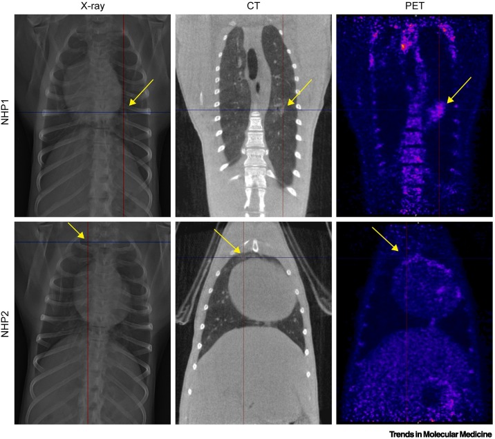
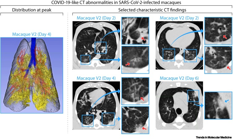
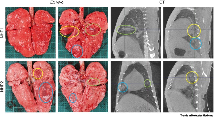
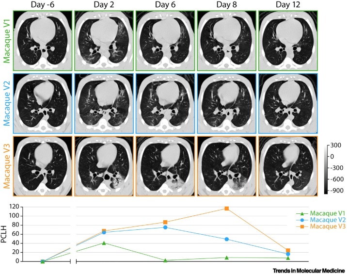
Chest X-ray (CXR), computed tomography (CT), and positron emission tomography-computed tomography (PET-CT) are noninvasive imaging techniques widely used in human and veterinary pulmonary research and medicine. These techniques have recently been applied in studies of severe acute respiratory syndrome coronavirus 2 (SARS-CoV-2)-exposed non-human primates (NHPs) to complement virological assessments with meaningful translational readouts of lung disease. Our review of the literature indicates that medical imaging of SARS-CoV-2-exposed NHPs enables high-resolution qualitative and quantitative characterization of disease otherwise clinically invisible and potentially provides user-independent and unbiased evaluation of medical countermeasures (MCMs). However, we also found high variability in image acquisition and analysis protocols among studies. These findings uncover an urgent need to improve standardization and ensure direct comparability across studies.

Keywords: COVID-19; SARS-CoV-2; imaging; lung; non-human primate.


Conflict of interest statement

Declaration of interests The authors declare no competing interests.

Figures

Figure 1
Figure 1
Comparison of chest X-ray (CXR), computed tomography (CT), and positron emission tomography–computed tomography (PET-CT) imaging. To compare results in non-human primate (NHP) lungs after severe acute respiratory syndrome coronavirus 2 (SARS-CoV-2) exposure, images on the same row originate from the same animal and were obtained on the same day (day 4 post-exposure). For the animal in the upper row, the lesion located in the middle part of the left lung (marked by the crosshairs and arrows) is clearly visible using all three imaging modalities. By contrast, alterations in lung density for the animal shown in the bottom row could only be clearly distinguished from healthy lung using CT (marked by the crosshairs and arrows) but are hardly visible on CXR and PET-CT. Adapted from [9].
Figure 2
Figure 2
Common lung abnormalities detected by computed tomography (CT) of severe acute respiratory syndrome coronavirus 2 (SARS-CoV-2)-exposed macaques. Distribution of CT scan abnormalities in a 3D reconstruction image of a SARS-CoV-2-exposed macaque at peak disease, day 4 (left panel: blue, airways; gray, normal lung; red, vessels; yellow, imaging abnormalities). Selected characteristic abnormalities include: (i) peri-bronchial consolidation in the left accessory lobe (top-middle panel day 2: top inset, red arrow); (ii) posterior ground glass opacities (GGOs) with reticulation in the posterior right lung (top-middle panel day 2: bottom inset, red arrow); (iii) bilateral posterior GGOs with reticulation (top-right panel day 2: top and bottom insets, red arrows); (iv) GGOs with superimposed crazy-paving pattern (interlobular septal thickening) in right posterior lung (bottom-middle panel day 4: top inset, red arrow) and mixed GGOs with pleural-based consolidation in left posterior lung (bottom-middle panel day 4: bottom inset, red arrow); and (v) pleural-based mixed GGOs and consolidation (bottom-right panel day 6: inset, blue arrow). Adapted from [8]. Abbreviation: COVID-19, coronavirus disease 2019.
Figure 3
Figure 3
Lung abnormalities detected by computed tomography (CT) matched with gross pathology. Obtained just before euthanasia (day 7 post-exposure), these images were matched to gross pathology photographs. Lesions found reflecting the same location are marked with similarly colored circles. Adapted from [61]. Abbreviation: NHP, non-human primate.
Figure 4
Figure 4
Data analysis of lung abnormalities detected by computed tomography (CT) of severe acute respiratory syndrome coronavirus 2 (SARS-CoV-2)-exposed macaques via percent change in lung hyperdensity (PCLH). Top: representative axial CT images in three SARS-CoV-2-exposed macaques for each indicated study day (D). The gray scale represents radiodensity in Hounsfield units (HU). Bottom: percentage change in volume of lung hyperdensity (PCLH) measured over time in the same SARS-CoV-2 inoculated macaques shown in the upper panel. Adapted from [8].

Similar articles

Cited by

References

    1. Rubin G.D., et al. The role of chest imaging in patient management during the COVID-19 pandemic: a multinational consensus statement from the Fleischner Society. Radiology. 2020;296:172–180. - PMC - PubMed
    1. Pan F., et al. Time course of lung changes at chest CT during recovery from coronavirus disease 2019 (COVID-19) Radiology. 2020;295:715–721. - PMC - PubMed
    1. Ai T., et al. Correlation of chest CT and RT-PCR testing for coronavirus disease 2019 (COVID-19) in China: a report of 1014 cases. Radiology. 2020;296:E32–E40. - PMC - PubMed
    1. Cleary S.J., et al. Animal models of mechanisms of SARS-CoV-2 infection and COVID-19 pathology. Br. J. Pharmacol. 2020;177:4851–4865. - PMC - PubMed
    1. Zamorano Cuervo N., Grandvaux N. ACE2: evidence of role as entry receptor for SARS-CoV-2 and implications in comorbidities. eLife. 2020;9 - PMC - PubMed

Publication types