. 2021 Dec 9:12:780727.
doi: 10.3389/fimmu.2021.780727. eCollection 2021.

NFAT5 Controls the Integrity of Epidermis

Affiliations

NFAT5 Controls the Integrity of Epidermis

Khalid Muhammad et al. Front Immunol. .

Abstract

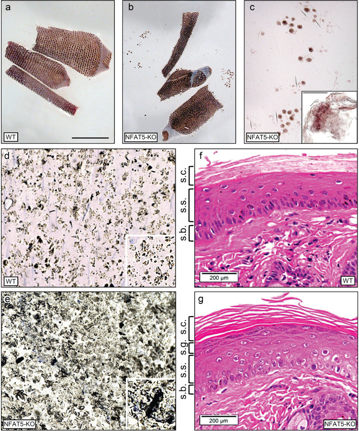
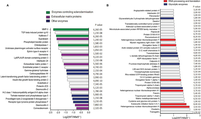
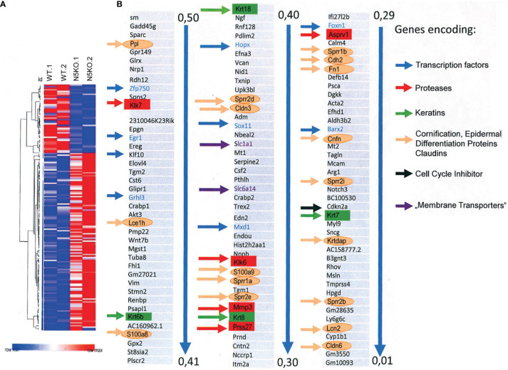
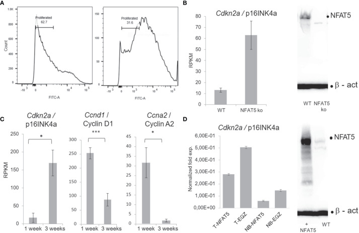
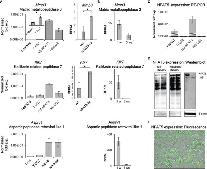
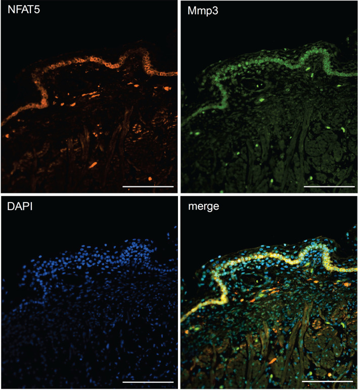
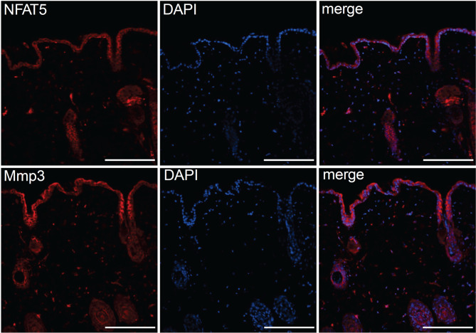
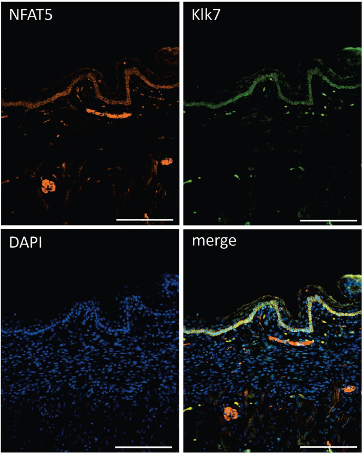
The skin protects the human body against dehydration and harmful challenges. Keratinocytes (KCs) are the most abundant epidermal cells, and it is anticipated that KC-mediated transport of Na+ ions creates a physiological barrier of high osmolality against the external environment. Here, we studied the role of NFAT5, a transcription factor whose activity is controlled by osmotic stress in KCs. Cultured KCs from adult mice were found to secrete more than 300 proteins, and upon NFAT5 ablation, the secretion of several matrix proteinases, including metalloproteinase-3 (Mmp3) and kallikrein-related peptidase 7 (Klk7), was markedly enhanced. An increase in Mmp3 and Klk7 RNA levels was also detected in transcriptomes of Nfat5-/- KCs, along with increases of numerous members of the 'Epidermal Differentiation Complex' (EDC), such as small proline-rich (Sprr) and S100 proteins. NFAT5 and Mmp3 as well as NFAT5 and Klk7 are co-expressed in the basal KCs of fetal and adult epidermis but not in basal KCs of newborn (NB) mice. The poor NFAT5 expression in NB KCs is correlated with a strong increase in Mmp3 and Klk7 expression in KCs of NB mice. These data suggests that, along with the fragile epidermis of adult Nfat5-/- mice, NFAT5 keeps in check the expression of matrix proteases in epidermis. The NFAT5-mediated control of matrix proteases in epidermis contributes to the manifold changes in skin development in embryos before and during birth, and to the integrity of epidermis in adults.

Keywords: Mmp3; NFAT5; epidermis; kallikrein 7; keratinocytes; matrix proteases; skin.


Conflict of interest statement

The authors declare that the research was conducted in the absence of any commercial or financial relationships that could be construed as a potential conflict of interest.

Figures

Figure 1
Figure 1
NFAT5 ablation changes the integrity and morphology of murine epidermis. (A, B) Preparations of skin from 129sv WT (A) and 129sv Nfat5-/- mice (B, C). In (C), skin sheets released from tail skin of Nfat5-/- mice are presented at larger magnification. Bar= 100 μM. (D, E) Tape strips from the tails of WT (D) and Nfat5-/- mice (E). One representative strip of more than ten is shown. (F, G) Representative H&E stains of skin sections from tails of WT (F) and Nfat5-/- mice (G). s.c., stratum corneum; s.g., stratum granulosum; s.s., stratum spinosum; s.b., stratum basale.
Figure 2
Figure 2
Proteins that were secreted by isolated tail KCs within 24 h of culture. (A) Proteins that were 2-fold and more secreted by 129sv Nfat5-/- than 129sv WT KCs. (B) Proteins that were at least 2-fold less secreted by Nfat5-/- than WT KCs. Data of two independent experiments each analyzed in triplicates by LC-MS were compiled.
Figure 3
Figure 3
Ablation of NFAT5 changed gene expression in cultivated KCs. (A) Heat map of 168 genes that were changed at least twofold in 129sv Nfat5-/- compared to 129sv WT KCs. (B) Compilation of 121 genes that were expressed in 2-fold more copies in Nfat5-/- KCs. Nine genes encoding TFs are shown in blue and indicated by blue arrows. Five protease genes are boxed and shown in red, four keratin genes are shown in green boxes and by green arrows. The gene coding for the cell cycle inhibitor Cdkn2a is shown in black, genes encoding membrane transporters are shown violet, and the numerous genes encoding Epidermal Differentiation Complex (EDC) proteins, claudins and other ‘cornification` proteins are encircled and presented in orange.
Figure 4
Figure 4
NFAT5 supports the proliferation of KCs. (A) Measurement of KC proliferation cultivated for one week (left) or three weeks (right) in CFSE assays. (B) Increase of p16INK4a RNA levels in KCs upon NFAT5 ablation. The western blot (right) confirms the absence of NFAT5 expression in KCs isolated from Nfat5-/- mice. (C) Expression of cell cycle inhibitor p16INK4a and of cyclins D1 and A2 in KCs cultivated for one or three weeks. Next-generation-sequencing (NGS) data of four independent batches of KCs cultured for 1 or 3 weeks are shown. (D) Decrease in p16INK4a RNA levels in KCs isolated from the tails of adult (T) or from newborn mice (NB). The western blot illustrates increased NFAT5 levels in tail KCs that have been transduced with a retrovirus expressing NFAT5. The KCs in (C, D) were cultivated for 1 week. (***p<0.0001, **p<0.001 and *p<0.05).
Figure 5
Figure 5
NFAT5 affects the expression of Mmp3, Klk5 and Asprv1 protease genes in KCs. (A) Left panels, results of qRT-PCR assays showing the effect of NFAT5 on the expression of Mmp3, Klk7 and Asprv1 genes upon transduction of KCs obtained from the tails of adult ice (T) or from the skin of newborn (NB) mice. Right panels, effect of NFAT5 ablation on Mmp3 and Klk7 expression. Results of 3 independent NGS assays are shown. (B) Expression of protease genes in KCs cultured for 1 or 3 weeks (ws). NGS data of three independent batches of KCs cultured for 1 or 3 ws are shown. (C–E) Detection of NFAT5 expression upon transduction of KCs. Cultured KCs were transduced with retroviruses expressing NFAT5, or with an ‘empty’ EGZ virus. (C) Detection of human NFAT5 RNA by RT-PCR assays in tail (T) and NB KCs upon transduction. (D) Western blots showing the expression of NFAT5 upon transduction of KCs from newborn and tails of adult mice. (E) Fluorescence microscopy of NFAT5-GFP expression in transduced adult KCs. (*p<0.05).
Figure 6
Figure 6
Immunofluorescence of sections through the skin of a human fetus. Sections were stained with Abs against NFAT5 (ab110995, Abcam) and Mmp3 (AF513, Novus Biologicals) and counterstained with DAPI as indicated. Length of the bars: 100 μm.
Figure 7
Figure 7
Immunofluorescence of serial sections through the back skin of adult mice. Sections were stained with Abs against NFAT5 (ab110995, Abcam) and Mmp3 (ab53015 (Abcam) and counterstained with DAPI as indicated. Length of the bars: 100 μm.
Figure 8
Figure 8
Immunofluorescence of sections through the skin of a human fetus. Sections were stained with Abs against NFAT5 (ab110995, Abcam) and Klk7 (Biotechn/R+D Systems, No.: AF2624) and counterstained with DAPI as indicated. Length of the bars: 100 μm.

References

    1. Proksch E, Brandner JM, Jensen JM. The Skin: An Indispensable Barrier. Exp Dermatol (2008) 17(12):1063–72. doi: 10.1111/j.1600-0625.2008.00786.x - DOI - PubMed
    1. Hofmeister LH, Perisic S, Titze J. Tissue Sodium Storage: Evidence for Kidney-Like Extrarenal Countercurrent Systems? Pflugers Arch (2015) 467(3):551–8. doi: 10.1007/s00424-014-1685-x - DOI - PMC - PubMed
    1. Jantsch J, Schatz V, Friedrich D, Schroder A, Kopp C, Siegert I, et al. . Cutaneous Na+ Storage Strengthens the Antimicrobial Barrier Function of the Skin and Boosts Macrophage-Driven Host Defense. Cell Metab (2015) 21(3):493–501. doi: 10.1016/j.cmet.2015.02.003 - DOI - PMC - PubMed
    1. Jobin K, Stumpf NE, Schwab S, Eichler M, Neubert P, Rauh M, et al. . A High-Salt Diet Compromises Antibacterial Neutrophil Responses Through Hormonal Perturbation. Sci Transl Med (2020) 12(536):1-13. doi: 10.1126/scitranslmed.aay3850 - DOI - PubMed
    1. Brooke MA, Nitoiu D, Kelsell DP. Cell-Cell Connectivity: Desmosomes and Disease. J Pathol (2012) 226(2):158–71. doi: 10.1002/path.3027 - DOI - PubMed

Publication types

Substances