. 2021 Dec 17;11(24):15051-15067.
doi: 10.1021/acscatal.1c03760. Epub 2021 Dec 1.

Intraparticle Kinetics Unveil Crowding and Enzyme Distribution Effects on the Performance of Cofactor-Dependent Heterogeneous Biocatalysts

Affiliations

Intraparticle Kinetics Unveil Crowding and Enzyme Distribution Effects on the Performance of Cofactor-Dependent Heterogeneous Biocatalysts

Eleftheria Diamanti et al. ACS Catal. .

Abstract

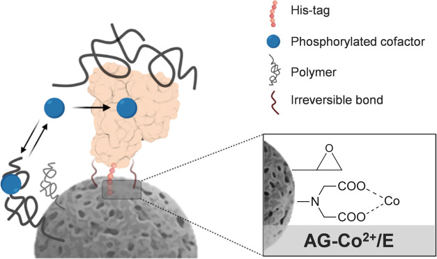
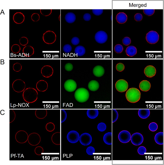
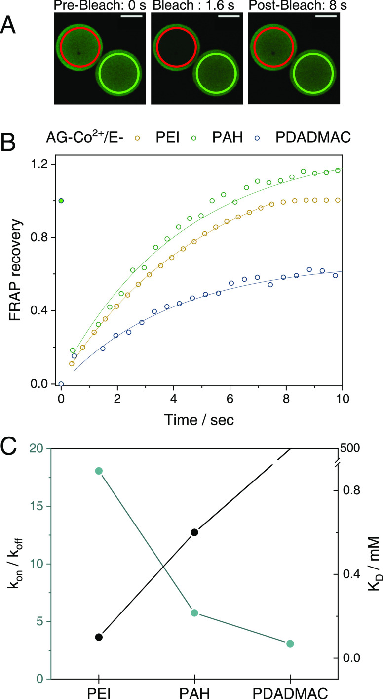
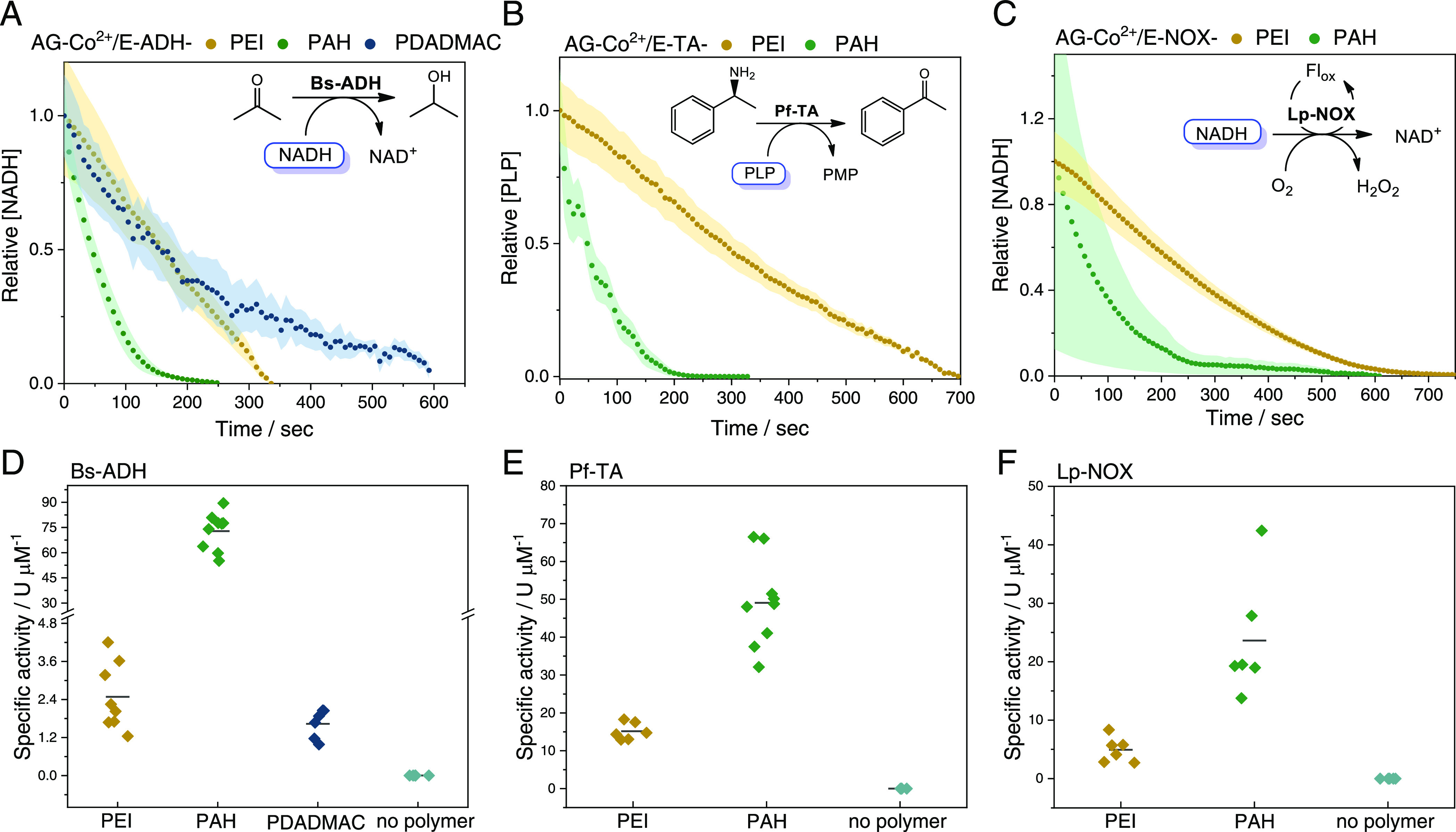
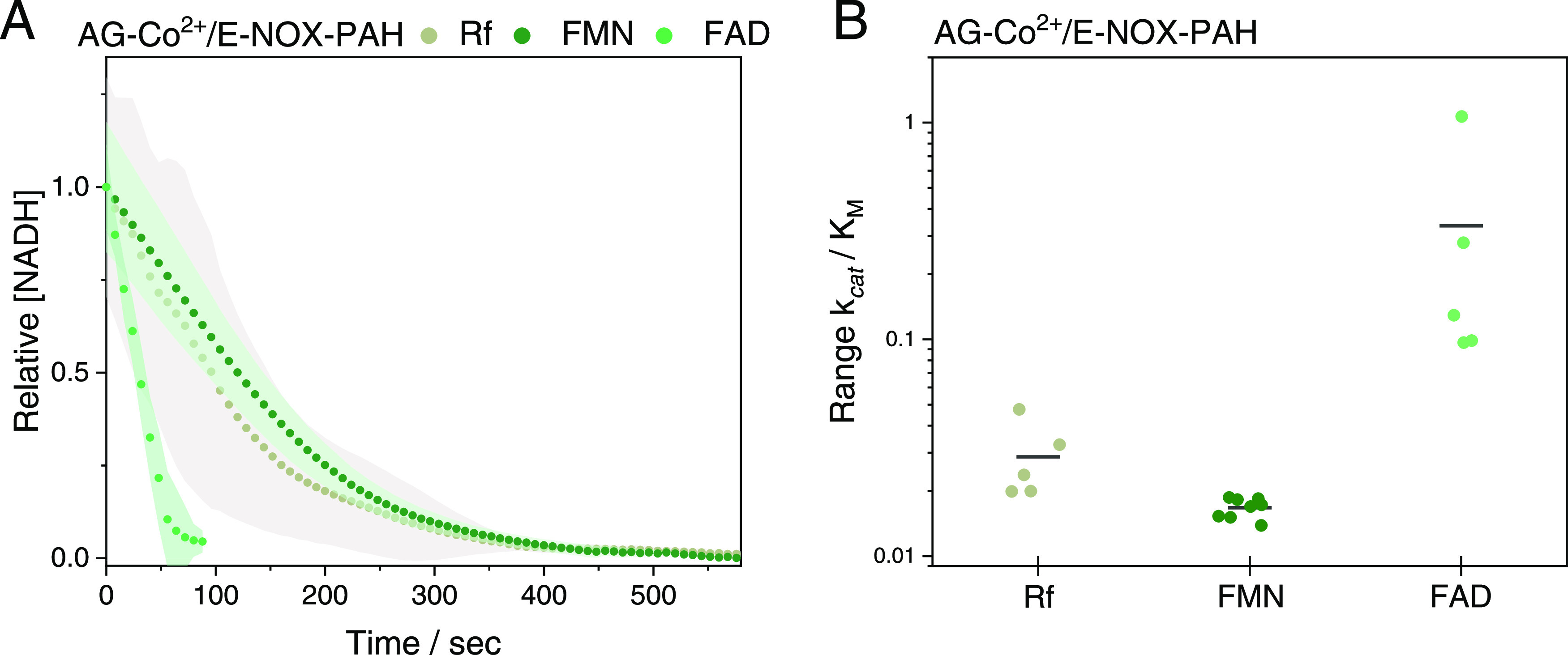
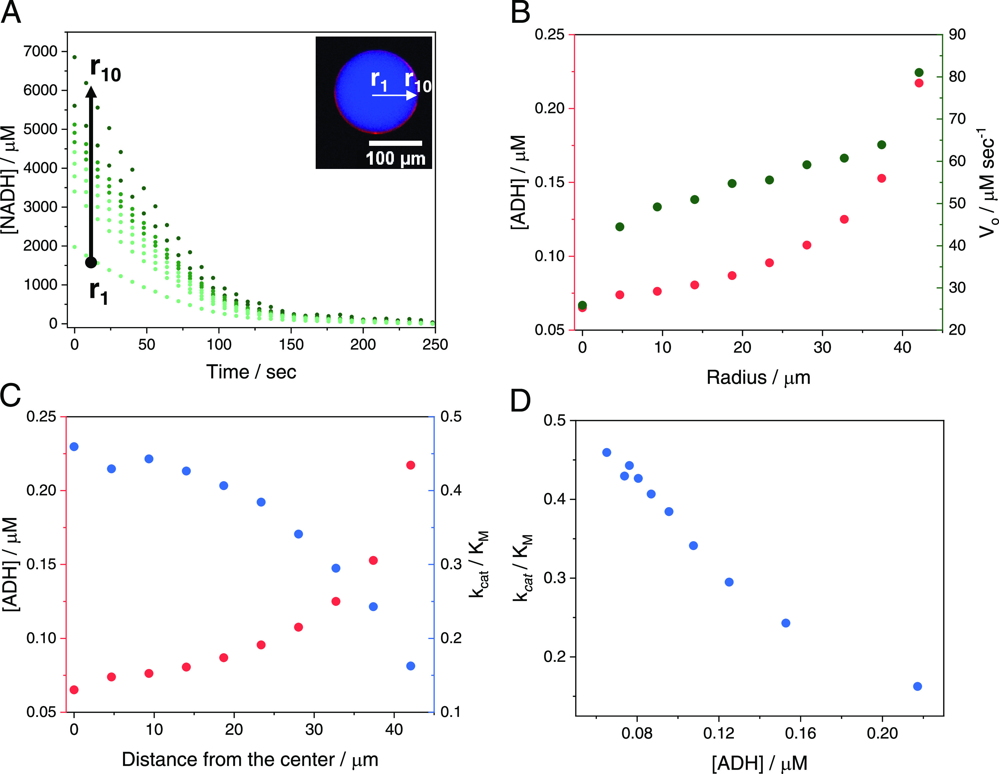
Multidimensional kinetic analysis of immobilized enzymes is essential to understand the enzyme functionality at the interface with solid materials. However, spatiotemporal kinetic characterization of heterogeneous biocatalysts on a microscopic level and under operando conditions has been rarely approached. As a case study, we selected self-sufficient heterogeneous biocatalysts where His-tagged cofactor-dependent enzymes (dehydrogenases, transaminases, and oxidases) are co-immobilized with their corresponding phosphorylated cofactors [nicotinamide adenine dinucleotide phosphate (NAD(P)H), pyridoxal phosphate (PLP), and flavin adenine dinucleotide (FAD)] on porous agarose microbeads coated with cationic polymers. These self-sufficient systems do not require the addition of exogenous cofactors to function, thus avoiding the extensive use of expensive cofactors. To comprehend the microscopic kinetics and thermodynamics of self-sufficient systems, we performed fluorescence recovery after photobleaching measurements, time-lapse fluorescence microscopy, and image analytics at both single-particle and intraparticle levels. These studies reveal a thermodynamic equilibrium that rules out the reversible interactions between the adsorbed phosphorylated cofactors and the polycations within the pores of the carriers, enabling the confined cofactors to access the active sites of the immobilized enzymes. Furthermore, this work unveils the relationship between the apparent Michaelis-Menten kinetic parameters and the enzyme density in the confined space, eliciting a negative effect of molecular crowding on the performance of some enzymes. Finally, we demonstrate that the intraparticle apparent enzyme kinetics are significantly affected by the enzyme spatial organization. Hence, multiscale characterization of immobilized enzymes serves as an instrumental tool to better understand the in operando functionality of enzymes within confined spaces.


Conflict of interest statement

The authors declare no competing financial interest.

Figures

Figure 1
Figure 1
Schematic illustration of porous agarose microbeads functionalized with cobalt chelates and epoxy groups for the site-selective irreversible enzyme immobilization and the assembly of the cationic polymers for the subsequent cofactor co-immobilization.
Figure 2
Figure 2
Spatial organization of enzymes and phosphorylated cofactors inside porous microbeads. Fluorescence microscopy images of (A) rhodamine B (RhB)-labeled Bs-ADH (red channel, λex: 561 nm) co-immobilized with fluorescent NADH (blue channel, λex: 405 nm), (B) RhB-labeled Lp-NOX (red channel, λex: 561 nm) co-immobilized with fluorescent FAD (green channel, λex: 488 nm) and (C) RhB-labeled Pf-TA (red channel, λex: 561 nm) co-immobilized with fluorescent PLP (blue channel, λex: 405 nm), on AG-Co2+/E-PAH carriers.
Figure 3
Figure 3
Fluorescence recovery after photobleaching (FRAP) analysis to study the intraparticle diffusion of cofactors. (A) Confocal fluorescence images before, during, and after photobleaching of the fluorescent FAD (green channel, λex: 488 nm) adsorbed on AG-Co2+/E carriers coated with PEI. The red circular region of interest (ROI, 93 μm) represents the photobleached area, and the green ROI represents the nonbleached area of the same size, used as a control. Scale bar, 50 μm. (B) FRAP normalized curves of FAD recovery when adsorbed to the different polycations PEI (yellow), PAH (green), and PDADMAC (blue). Dots represent the experimental data, while the solid line corresponds to the full reaction–diffusion model fitting (see the Materials and Methods section). (C) Pseudoequilibrium constant kon/koff calculated from FRAP analysis as a function of the dissociation constant KD calculated from Langmuir adsorption Isotherms (Table 1). KD data point for PDADMAC (not visible) is ≫500 according to its linear adsorption isotherm (see Figure S4).
Figure 4
Figure 4
Single-particle activity of different enzyme/cofactor pairs co-immobilized on AG-Co2+/E coated with different polycations: PEI (yellow), PAH (green), and PDADMAC (blue). (A–C) Single-particle normalized mean time courses of the relative cofactor concentration. Time data points are obtained from the mean value of 10 microbeads with the standard deviation depicted in shadows of the same color. (D–F) Single-particle specific activity of different immobilized systems. Each data point represents the specific activity of one single bead. Specific activity is defined as the activity units per enzyme concentration (U μM–1). The activity unit (U) is defined as the concentration of the cofactor consumed per second (μM s–1). (A and D) Bs-ADH co-immobilized with NADH using acetone as an exogenous substrate, (B and E) Pf-TA co-immobilized with PLP using rac-phenylethylamine as a substrate, and (C and F) Lp-NOX co-immobilized with NADH using riboflavin as a flavin cofactor. Insets (A–C) reaction schemes of each biocatalyst.
Figure 5
Figure 5
Single-particle reaction of Lp-NOX co-immobilized with NADH on AG-Co2+/E-PAH in the presence of different flavins: riboflavin (Rf, gray), FMN (dark green), and FAD (light green). (A) Single-particle normalized mean time courses of the relative NADH concentration. Time data points are obtained from the mean value of 10 microbeads with the standard deviation depicted in shadows of the same color. (B) Apparent catalytic efficiency (kcat/KM) of Lp-NOX co-immobilized with NADH on AG-Co2+/E-PAH in the presence of Rf, FMN, and FAD, as flavin cofactors. Each data point represents the apparent catalytic efficiency toward the confined NADH in one single bead.
Figure 6
Figure 6
Effect of microbead radius and enzyme concentration on the apparent Michaelis–Menten kinetics of Bs-ADH co-immobilized with NADH on AG-Co2+/E-PAH. (A) Apparent kcat/KM toward NADH as a function of the microbead radius. (B) Apparent kcat/KM toward NADH as a function of the enzyme concentration immobilized on one single microbead. (C) Apparent kcat and (D) apparent KM toward NADH versus the concentration of immobilized Bs-ADH. Each data point represents the corresponding apparent kinetic parameter toward the confined NADH in one single microbead. For statistical measurements, we performed linear regression (OriginLab) on each scatterplot and analysis of variance (ANOVA) statistical analysis to derive Pearson′s correlation coefficient (r) and P-values, respectively. Statistical analysis of (A), (B), (C), and (D) resulted in r = 0.7 (p < 0.005), r = −0.9 (p < 0.005), r = −0.6 (p < 0.005), and r = 0.8 (p < 0.005), respectively.
Figure 7
Figure 7
Intraparticle kinetic studies of Bs-ADH co-immobilized with NADH on AG-Co2+/E-PAH. (A) Intraparticle time courses of NADH oxidation in the presence of acetone at different positions of the radial intensity profile (r1: center, r10: outer surface of one single microbead). Top right inset: representation of the radial intensity profile in the merged fluorescence image of RhB-labeled Bs-ADH (red channel, λex: 523 nm) co-immobilized with fluorescent NADH (blue channel, λex: 365 nm). (B) Intraparticle enzyme concentration (left y-axis) and initial rate Vo (right y-axis) measured at different positions of the radial intensity profile. (C) Intraparticle enzyme concentration (left y-axis) and apparent kcat/KM (right y-axis) as determined from the time courses in panel (A) at different distances from the center of the microbead. (D) Intraparticle apparent kcat/KM as a function of the local Bs-ADH concentration within one single microbead.
Figure 8
Figure 8
Effect of spatial distribution on the single-particle and intraparticle kinetics of Bs-ADH co-immobilized with NADH on AG-Co2+/E-PAH. (A and B) Epifluorescence and confocal fluorescence (top right inset) merged images of RhB-labeled Bs-ADH (red channel, λex: 523 nm) immobilized with different spatial distributions, and co-immobilized with fluorescent NADH (blue channel, λex: 365 nm) on AG-Co2+/E-PAH carriers. (A) Bs-ADH immobilization in the presence of 50 mM imidazole resulting in patches at the outer surface of the microbead and (B) Bs-ADH immobilization in the presence of 200 mM imidazole resulting in a uniform distribution across the microbead, where r (white line) depicts the radial intensity profile position of one selected microbead. (C) Single-particle time courses of NADH oxidation in the presence of acetone using immobilized systems with different Bs-ADH spatial distributions; no imidazole (gray, see Figure 6A), imidazole 50 mM (yellow), and 200 mM imidazole (blue). Time data points are obtained from the mean value of 10 microbeads with the standard deviation depicted in shadows of the same color. The first and second phases of the biphasic time courses are represented with symbols I and II, respectively. (D) Initial rate and enzyme-specific activity calculated from Phase I of the time courses shown in panel C for each immobilized enzyme with the different spatial distribution. Specific activity is defined as the activity units per enzyme concentration (U μM–1). The activity unit (U) is defined as the concentration of the cofactor consumed per second (μM s–1). (E) Radial intensity profiles of the intraparticle enzyme concentration and intraparticle specific activity within one single microbead of the self-sufficient heterogeneous biocatalyst prepared in the presence of 50 mM imidazole. (F) Radial intensity profiles of the intraparticle enzyme concentration and intraparticle specific activity within one single microbead of the self-sufficient heterogeneous biocatalyst prepared in the presence of 200 mM imidazole.

References

    1. Di Cosimo R.; Mc Auliffe J.; Poulose A. J.; Bohlmann G. Industrial Use of Immobilized Enzymes. Chem. Soc. Rev. 2013, 42, 6437–6474. 10.1039/c3cs35506c. - DOI - PubMed
    1. Guisan J. M.; Bolivar J. M.; López-gallego F.. Immobilization of Enzymes and Cells; Guisan J. M.; Bolivar J. M.; López-Gallego F.; Rocha-Martín J., Eds.; Methods in Molecular Biology; Springer US: New York, NY, 2020; Vol. 2100. - PubMed
    1. Bommarius A. S.; Paye M. F. Stabilizing Biocatalysts. Chem. Soc. Rev. 2013, 42, 6534–6565. 10.1039/C3CS60137D. - DOI - PubMed
    1. Brena B.; González-Pombo P.; Batista-Viera F.. Immobilization of Enzymes: A Literature Survey. In Immobilization of Enzymes and Cells, 3rd ed.; Guisan J. M., Ed.; Methods in Molecular Biology; Humana Press: Totowa, NJ, 2013; Vol. 1051, pp 15–31. - PubMed
    1. Garcia-Galan C.; Berenguer-Murcia Á.; Fernandez-Lafuente R.; Rodrigues R. C. Potential of Different Enzyme Immobilization Strategies to Improve Enzyme Performance. Adv. Synth. Catal. 2011, 353, 2885–2904. 10.1002/adsc.201100534. - DOI