Review
. 2021 Dec 9:25:101742.
doi: 10.1016/j.jcot.2021.101742. eCollection 2022 Feb.

Imaging update in spinal tuberculosis

Affiliations
Review

Imaging update in spinal tuberculosis

Vijay Kubihal et al. J Clin Orthop Trauma. .

Abstract

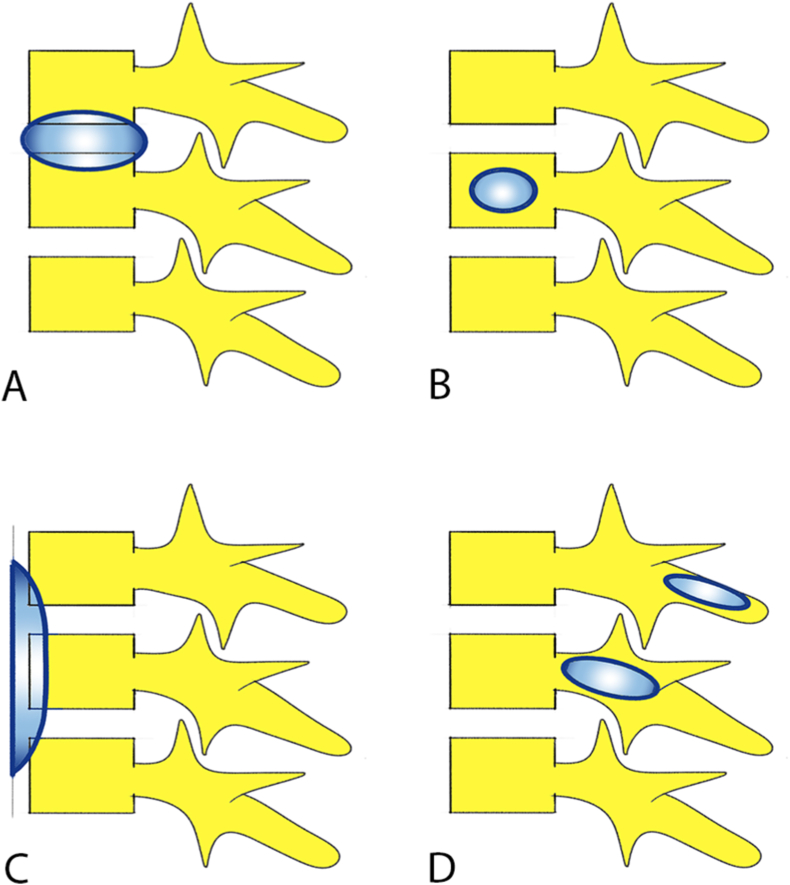
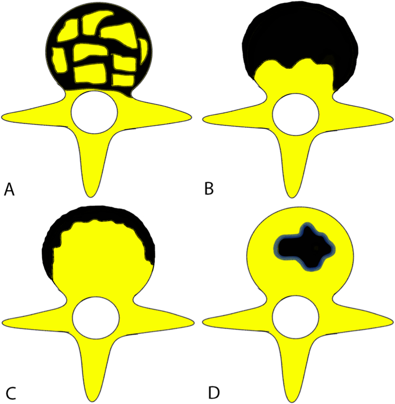
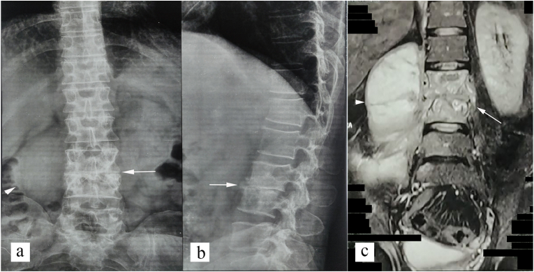
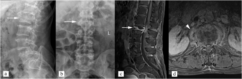
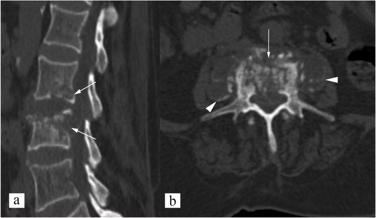
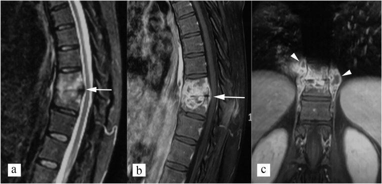
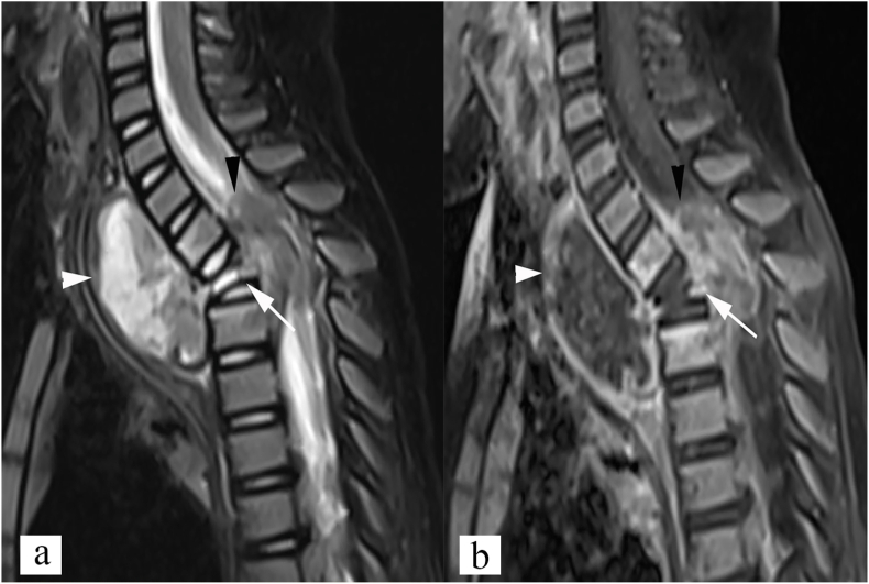
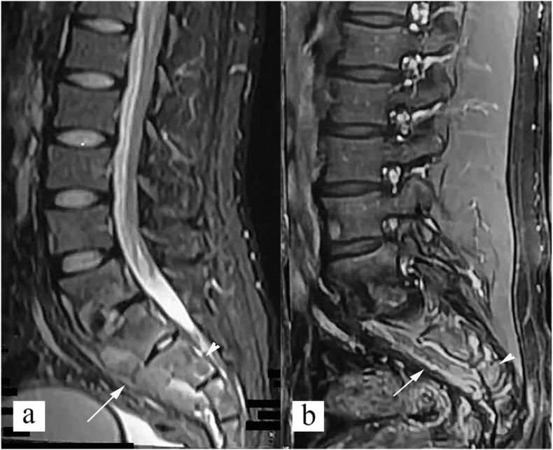
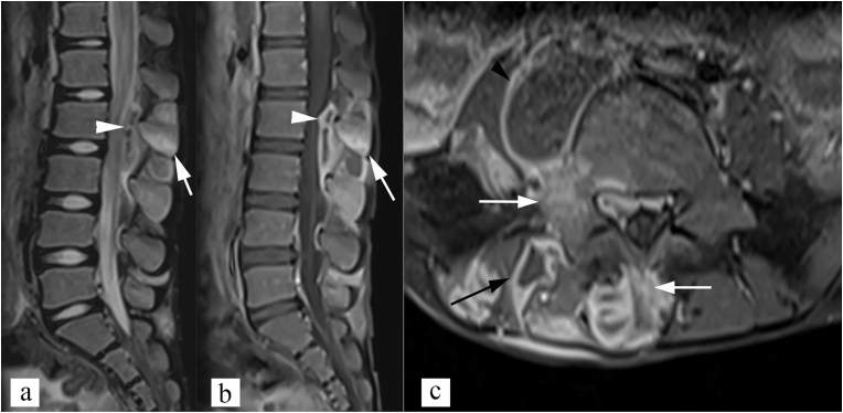
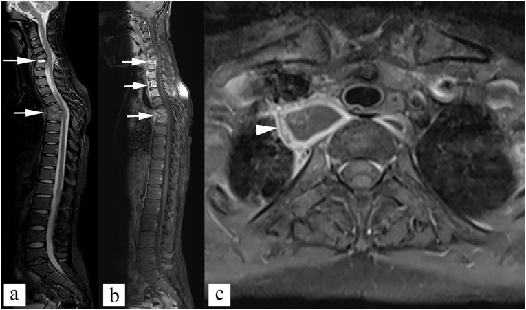
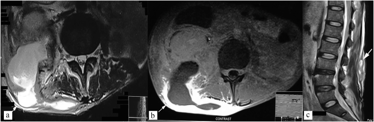
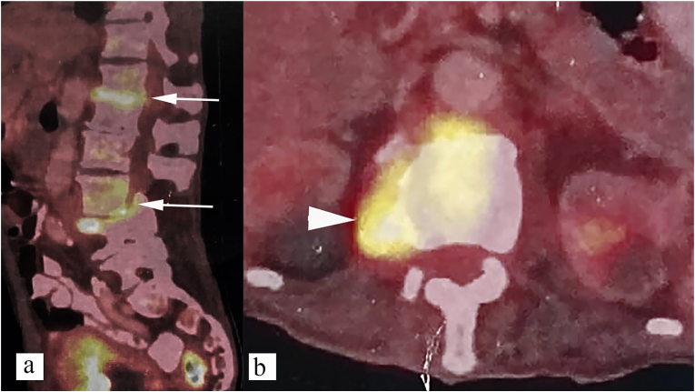
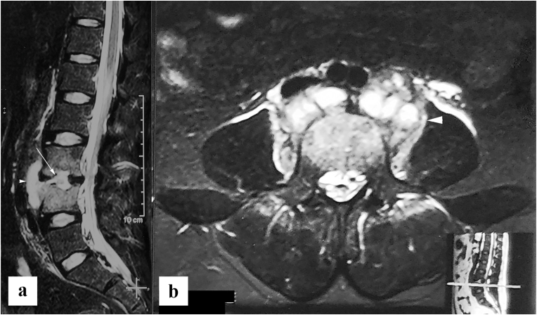
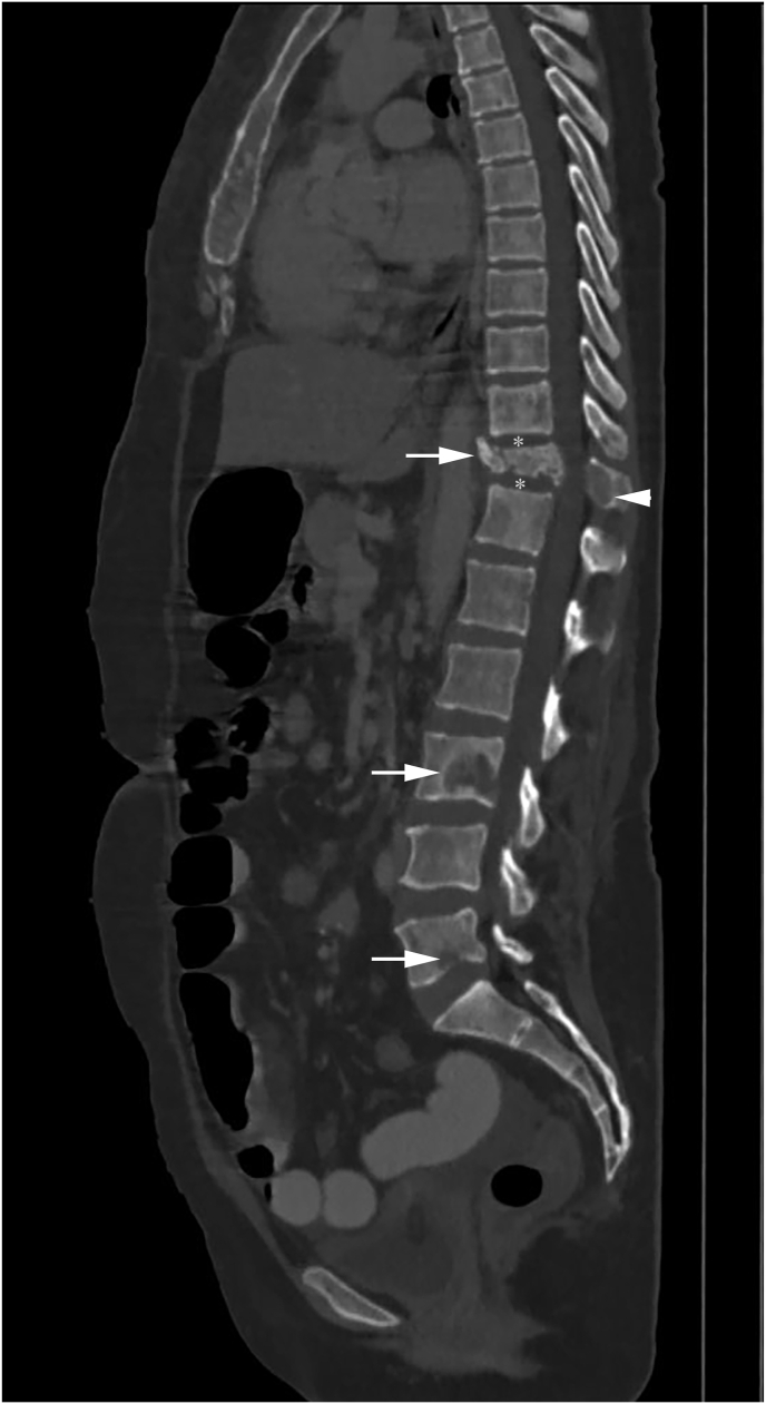
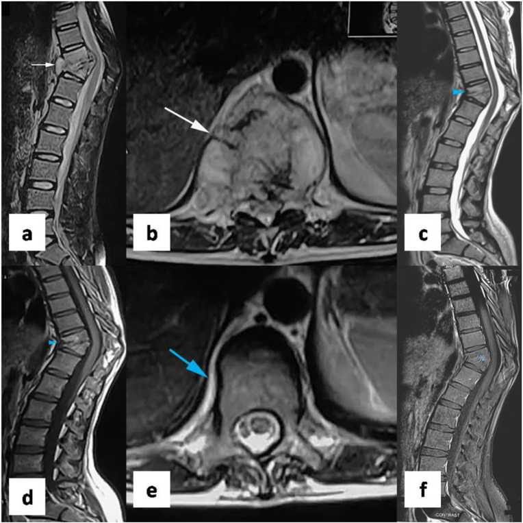
Tuberculosis is ancient disease known to mankind. Diagnosis and management of spinal tuberculosis has immensely improved in last few decades. Imaging, particularly MRI, plays important role in diagnosis of spinal tuberculosis and its complications. Four common imaging patterns of spinal tuberculosis include paradiscal type, central type, Anterior subligamentous type, and posterior type. Imaging also plays important role in differentiation of spinal tuberculosis from its mimics, particularly pyogenic spondylitis, and metastasis. Radiological interventions, such as CT guided vertebral biopsy, and percutaneous drainage of cold abscess, are commonly used in management of spinal tuberculosis. Monitoring of therapeutic response is often based on clinical evaluation and imaging. MRI is most common imaging modality used. Signs of healing include bony ankylosis, resolution of marrow edema, decrease in contrast enhancement, and fatty change with in bone marrow. PET CT is recently evaluated for response assessment with promising results. This review summarizes pathophysiology, clinical presentation, imaging features, radiological interventions, and response assessment in spinal tuberculosis.

Keywords: Imaging intervention; Response evaluation; Spinal infection imaging; Spinal tuberculosis.


Conflict of interest statement

None.

Figures

Fig. 1
Fig. 1
Patterns of vertebral involvement in spinal tuberculosis. (A: Paradiscal; B: Central; C: Anterior subligamentous; D: Posterior (neural arch) involvement).
Fig. 2
Fig. 2
Bone destruction types in spinal tuberculosis. (A: Fragmentary; B: Osteolytic; C: Subperiosteal; D: Localized destruction with sclerosed margins).
Fig. 3
Fig. 3
Radiographic features of spinal tuberculosis (paradiscal type). (a) anteroposterior and (b) lateral radiograph of lumbar spine shows loss of L3-L4 disc space, endplate erosions, lytic destruction of L3 and L4 vertebral bodies (white arrow), and right paravertebral soft tissue (white arrowhead). (c) coronal STIR image shows loss of L3-L4 disc space, marrow edema in L3, L4 (white arrow), and also L5 vertebral bodies, and right psoas abscess consistent with spinal tuberculosis (white arrowhead).
Fig. 4
Fig. 4
Radiographic features of spinal tuberculosis (central type). (a) Lateral and (b) Anteroposterior radiograph of lumbar spine shows isolated L2 vertebral involvement with concertina collapse of vertebral body (white arrow). No paravertebral soft tissue appreciated on radiographs. (c) sagittal post contrast T1 weighted image shows abnormal enhancement of L2 vertebral body (white arrow). (d) axial post contrast T1 weighted image shows enhancing paravertebral soft tissue (white arrowhead).
Fig. 5
Fig. 5
CT features of spinal tuberculosis (paradiscal type). (a) sagittal non-contrast CT image shows paradiscal end plate erosions, and vertebral body involvement of L2 and L3 vertebrae (white arrows). (b) axial non-contrast CT image shows fragmentary type bone destruction (white arrow), and paravertebral soft tissue (white arrowhead). Calcification is noted in paravertebral soft tissue, which is pathognomonic for spinal tuberculosis.
Fig. 6
Fig. 6
MRI findings in spinal tuberculosis, paradiscal type. (a) Sagittal STIR image, and (b) sagittal post contrast T1 weighted image shows reduced disc space, with inflammatory marrow edema of D10 and D11 vertebral bodies seen (white arrow). (c) Coronal post contrast T1 weighted image shows bilateral paravertebral peripherally enhancing cold abscesses (white arrowhead).
Fig. 7
Fig. 7
Central type of spinal tuberculosis. (a) STIR sagittal, and (b) post contrast T1 weighted sagittal MR images show inflammatory bone marrow edema involving D2 vertebral body (white arrow), epidural abscess compromising the spinal canal (black arrowhead), and prevertebral abscess (white arrowhead).
Fig. 8
Fig. 8
Anterior subligamentous type of spinal tuberculosis. Sagittal STIR (a), and post contrast T1 weighted (b) images show subligamentous abscess (white arrow) beneath the anterior longitudinal ligament along L5, and sacral vertebrae, causing scalloping of anterior vertebral margins (white arrow). In addition, inflammatory marrow edema also noted in L5, and sacral vertebral bodies, adjacent to the abscess (white arrowhead).
Fig. 9
Fig. 9
Posterior type of spinal tuberculosis. (a) Sagittal STIR image, and (b) sagittal post contrast T1 weighted image show inflammatory marrow edema in L2 and L3 spinous process (white arrow), with epidural abscess (white arrowhead). Note that vertebral bodies shows normal marrow signal. (c) axial post contrast T1 weighted image shows involvement of spinous process, and right lamina, and pedicle (white arrow) with right posterior paravertebral abscess (black arrow) and right psoas abscess (black arrowhead).
Fig. 10
Fig. 10
Multifocal disease. (a) Sagittal STIR image, and (b) sagittal post contrast T1 weighted image shows multiple cervical and upper dorsal vertebral body involvement (white arrows). Axial post contrast T1 weighted image shows paravertebral cold abscess (white arrowhead).
Fig. 11
Fig. 11
Atypical presentation - isolated cold abscess. Axial STIR (a), and T1 weighted post contrast (b) images show tubercular cold abscess in right posterior paraspinal location, posterior abdominal wall, and posterior pararenal space (white arrow). (c) Sagittal T2 weighted image shows normal vertebra marrow signal intensity.
Fig. 12
Fig. 12
18F-FDG PET CT features of spinal tuberculosis (paradiscal type). Paradiscal increased metabolic activity seen at D12-L1 and L3-L4 vertebral levels (a) (White arrow), right paravertebral soft tissue (b) (white arrowhead).
Fig. 13
Fig. 13
Pyogenic spondylitis (biopsy proven) in 30year old male patient with acute onset fever and back pain. (a) Sagittal, and (b) axial STIR images show reduced L3-L4 intervertebral disc space, with abnormally high T2 signal intensity with in the disc (white arrow), and thick irregular walled paraspinal abscess (white arrowhead). Bone marrow edema seen in L3 and L4 vertebral bodies with relatively preserved vertebral height.
Fig. 14
Fig. 14
Spinal metastasis in a patient with infiltrating ductal carcinoma of the breast. Sagittal CT bone window image shows lytic lesions involving multiple non-contiguous vertebrae (skip lesions) (white arrow) with metastatic compression fracture of the D11 vertebra body, and involvement of posterior elements (white arrowhead). Intervertebral disc spaces are maintained (white asterisk).
Fig. 15
Fig. 15
MRI features of healing. (a) sagittal STIR and (b) axial T2 weighted images show paradiscal involvement of D10-D11 vertebrae with wedge collapse of D10 and D11 vertebral bodies, inflammatory bone marrow edema, and paravertebral soft tissue (white arrow) consistent with active spinal tuberculosis. Follow up MRI was done after 8 months of antitubercular therapy. (c) Sagittal T2 weighted and (d) sagittal T1 weighted images of follow up MRI shows bony ankylosis with fatty change in bone marrow (blue arrowhead). (e) axial T2 weighted image of follow up MRI shows resolution of paravertebral soft tissue (blue arrow). (f) Sagittal T1 weighted post contrast images of follow up MRI shows no abnormal contrast enhancement (blue asterisk). Follow up MRI features are consistent with healing.

References

    1. Duraiswami P.K., Orth M., Tuli S.M. 5000 years of orthopaedics in India. Clin Orthop. 1971 Apr;75:269–280. PMID: 4929008. - PubMed
    1. Rajasekaran S., Soundararajan D.C.R., Shetty A.P., Kanna R.M. Spinal tuberculosis: current concepts. Global Spine J. 2018 Dec;8(4_suppl):96S–108S. doi: 10.1177/2192568218769053. - DOI - PMC - PubMed
    1. Tuli S.M. Historical aspects of Pott's disease (spinal tuberculosis) management. Eur Spine J Off Publ Eur Spine Soc Eur Spinal Deform Soc Eur Sect Cerv Spine Res Soc. 2013 Jun;22(Suppl 4):529–538. doi: 10.1007/s00586-012-2388-7. - DOI - PMC - PubMed
    1. Jain A.K. Tuberculosis of spine: research evidence to treatment guidelines. Indian J Orthop. 2016 Feb;50(1):3–9. doi: 10.4103/0019-5413.173518. - DOI - PMC - PubMed
    1. Garg D., Goyal V. Spinal tuberculosis treatment: an enduring bone of contention. Ann Indian Acad Neurol. 2020;23(4):441. doi: 10.4103/aian.AIAN_141_20. - DOI - PMC - PubMed

LinkOut - more resources