Review
. 2021 Dec 10:11:780264.
doi: 10.3389/fonc.2021.780264. eCollection 2021.

Osteosarcoma and Metastasis

Affiliations
Review

Osteosarcoma and Metastasis

Gaohong Sheng et al. Front Oncol. .

Abstract

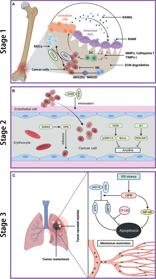
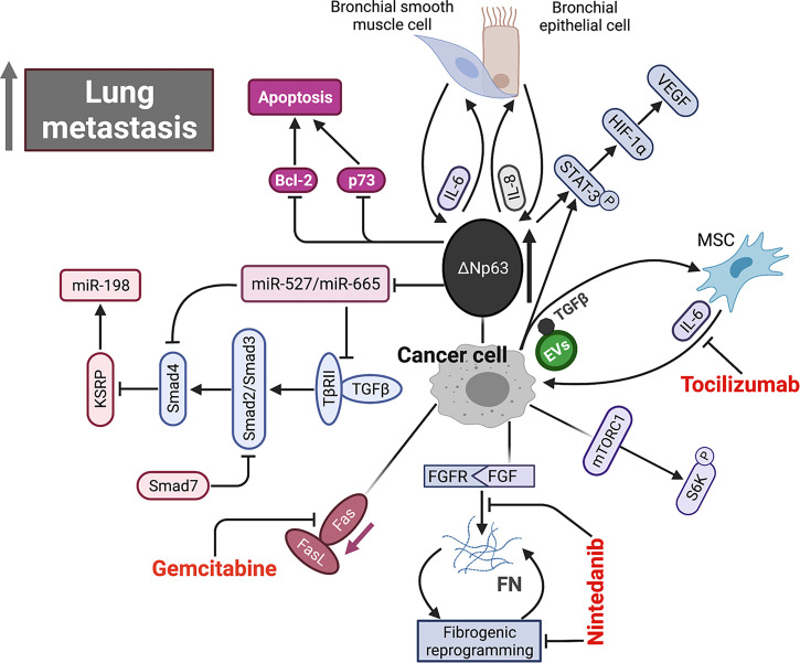
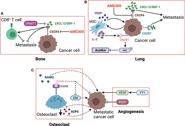
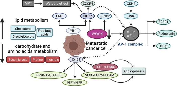
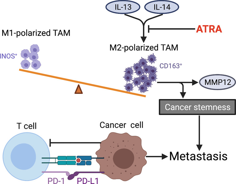
Osteosarcoma is the most common primary bone malignancy in adolescents. Its high propensity to metastasize is the leading cause for treatment failure and poor prognosis. Although the research of osteosarcoma has greatly expanded in the past decades, the knowledge and new therapy strategies targeting metastatic progression remain sparse. The prognosis of patients with metastasis is still unsatisfactory. There is resonating urgency for a thorough and deeper understanding of molecular mechanisms underlying osteosarcoma to develop innovative therapies targeting metastasis. Toward the goal of elaborating the characteristics and biological behavior of metastatic osteosarcoma, it is essential to combine the diverse investigations that are performed at molecular, cellular, and animal levels from basic research to clinical translation spanning chemical, physical sciences, and biology. This review focuses on the metastatic process, regulatory networks involving key molecules and signaling pathways, the role of microenvironment, osteoclast, angiogenesis, metabolism, immunity, and noncoding RNAs in osteosarcoma metastasis. The aim of this review is to provide an overview of current research advances, with the hope to discovery druggable targets and promising therapy strategies for osteosarcoma metastasis and thus to overcome this clinical impasse.

Keywords: metabolism; metastasis; microenvironment; noncoding RNAs; osteosarcoma.


Conflict of interest statement

The authors declare that the research was conducted in the absence of any commercial or financial relationships that could be construed as a potential conflict of interest.

Figures

Figure 1
Figure 1
Metastatic cascade of OS to the lung. (A) Stage 1: dissemination of metastatic OS cells from primary site. Cancer cells induce OB to secrete RNAKL, which binds to OC, and lead to bone resorption. The increase of MMPs and cathepsins while decrease of TIMPs cause ECM degradation. Cancer cells secret SDF-1 to recruit MSCs, which in turn promote tumor growth and metastasis by secreting CCL-5. NK cells kill cancer cell through the interaction between NKG2D receptor and NKG2D ligands (NKG2DL). (B) Stage 2: transfer of OS cells in blood. The interaction between uPAR and FPR1 enhances invasion of cancer cell and their entry into blood. RUNX2/OPN axis promotes adhesion of cancer cell to endothelia cell in lung. FASN and ID1 increase anoikis resistance in cancer cell by upregulating p-ERK1/2 and Bcl-xL and by activating PI3K/AKT pathway, respectively. (C) Stage 3: colonization of OS cells in lung. The ER stress-initiated UPR protects cancer cell from apoptosis by activating GRP78 and ATF6, as well as through NF-κB pathway. The mechanical restriction of circulating cancer cells within lung microvasculature partly accounts for the propensity of lung metastasis. Tumor-secreted vesicles reach the lung in advance and direct cancer cell to transfer to the lung. OS, osteosarcoma; OB, osteoblast; OC, osteoclast; RANK, receptor activator of NF-κB; RANKL, receptor activator of NF-κB ligand; MMPs, matrix metalloproteinases; TIMPs, tissue inhibitor of metalloproteinases; ECM, extracellular matrix; SDF-1, stromal cell-derived factor-1; MSCs, mesenchymal stem cells; CCL-5, C–C motif chemokine ligand 5; NKG2D, natural killer group 2 member D; uPAR, urokinase-type plasminogen activator; FPR1, formyl peptide receptor type 1; RUNX2, runt‐related transcription factor 2; OPN, osteopontin; FASN, fatty acid synthase; ID1, inhibitor of differentiation or DNA binding; ERK, extracellular signal-regulated kinase; Bcl-xL, B-cell lymphoma-extra large; ER, endoplasmic reticulum; UPR, unfolded protein response; GRP78, glucose-regulated protein 78KD; ATF6, activating transcription factor 6; IT-139, a novel small molecule that inhibit GRP78.
Figure 2
Figure 2
Schematic representation of signaling pathways within microenvironment underlying OS metastasis. Aberrant overexpression of ΔNp63 in cancer cell directly drives feed-forward amplification of IL-6 and IL-8 production by the interactions between cancer cell and both primary bronchial epithelial cell and bronchial smooth muscle cell. ΔNp63 overexpression leads to elevated phosphorylation of STAT-3, which further activates HIF-1α/VEGF axis. High expression of ΔNp63 promotes cancer cell survival by inhibiting Bcl-2 and p73-depedent apoptosis. In addition, ΔNp63 represses miR-527 and miR-665, leading to the upregulation of two TGF-β effectors, Smad4 and TβRII, which inhibits anti-metastasis miR-198 by suppressing its regulatory factor, KSRP. FGF signaling initiates and FN signaling sustains fibrotic reprogramming. Nintedanib targets the pan FGFR-FN axis to inhibit OS lung metastasis. Fas-negative OS cells are selected during metastasis by evading elimination in lung where Fas ligand (FasL) is constitutively expressed. Gemcitabine appears to be a promising agent by upregulating Fas expression. EVs secreted by OS cells selectively incorporate a membrane-associated form of TGF-β and induce IL-6 production by MSCs, which in turn promotes OS progression. IL-6, interleukin-6; IL-8, interleukin-8; STAT-3, signal transducer and activator of transcription 3; HIF-1α, hypoxia-inducible factor 1α; VEGF, vascular endothelial growth factor; TGF-β, tumor growth factor β; FGF, fibroblast growth factor; FN, fibronectin; EVs, extracellular vesicle.
Figure 3
Figure 3
The role of osteoclast and CXCR/CXCL axis in OS metastasis. (A) Highly expressed CXCL12. The CXCR4/CXCL12 (SDF-1) interaction is critical for OS metastasis in the lung, which is further strengthened by MSC via secreting VEGF. MSC-derived IL-8 induces OS cell anoikis resistance by activating CXCR1/Akt signaling. Another receptor CXCR7 expressed on OS cells promotes lung metastasis and enhances the malignancy activity of CXCR4. (B) RANK-Fc binds to RANK as a potent RANKL antagonist to inhibit osteoclast formation and activity, which can reduce OS metastasis, partly by suppressing ERK. Controversially, metastasis-competent OS cells induce loss of ACP5+ osteoclasts, which in turn enhances metastasis. Herein, we used dotted lines to indicate this contradiction. VEGF exhibits prometastatic effects on OS cells while PEDF shows the opposite by regulating angiogenesis. (C) In primary bone site, OS epigenetically downregulates CXCL12 expression by DNMT1, impairs cytotoxic T-cell homing to the tumor site, and this chemokine gradient of CXCL12 drives the metastasis of OS cells to the lung. ACP5/TRAP, osteoclast-specific tartrate-resistant acid phosphatase 5; PEDF, pigment epithelium-derived factor; YY1, Yin Yang 1 protein; CXCL12, C–X–C motif chemokine 12; DNMT1, DNA methyltransferase 1; CXCR4, chemokine receptor 4; AMD3100, CXCR4 antagonist.
Figure 4
Figure 4
Metabolic reprogramming during OS metastasis. MPT promotes Warburg effect in OS cells by suppressing mitochondrial function. The serum metabolic profile of lung metastasis shows lowered carbohydrate and amino acid metabolism but an elevated lipid metabolism. YB-1 contributes to metastasis by translational activation of EMT and HIF-1α, which then induces CXCR4 expression. Cyr61 enhances the metastatic potential of OS cells through multiple signaling pathways, including PI-3K/Akt/GSK3β, IGF1/IGFR, and angiogenesis-associated signaling (increased VEGF, FGF2, PECAM and reduced TSP-1, SPARC). WWOX maintains mitochondrial respiration and inhibits Warburg effect by physical interaction with HIF-1α. WWOX also suppresses c-Jun activity by physical association while CDH4 overexpression activates c-Jun via the JNK pathway. AP-1 is a transcriptional complex, mainly composed of c-Jun and c-Fos, which promotes metastatic potential by upregulating several downstream effectors, including FGFR1, podoplanin, and TGFβ. The dotted lines indicate the mechanistic study is performed in melanoma cells, that is, HIF-1α interacts with AP-1, which then binds to AP-1-binding motif within the Cyr61 promoter and induces Cyr61 expression. MPT, mitochondrial permeability transition; YB-1, Y-box binding protein 1; EMT, epithelial-to-mesenchymal transition; Cyr61, cysteine-rich protein 61; IGF-1, insulin-like growth factor 1; FGF2, fibroblast growth factor 2; PECAM, platelet endothelial cell adhesion molecule; TSP-1, thrombospondin-1; SPARC, secreted protein acidic and rich in cysteine; WWOX, WW domain-containing oxidoreductase; CDH4, cadherin-4; AP-1, activating protein-1; FGFR1, fibroblast growth factor receptor 1.
Figure 5
Figure 5
Tumor immune microenvironment characteristics within OS metastasis. The imbalance of M1 (INOS+)/M2 (CD163+)-polarized TAMs in favor of M2 subtype is observed in metastatic OS. ATRA suppresses IL-13-induced secretion of MMP12 from M2-polarized macrophages and also weakens cancer stemness by preventing M2 polarization of TAMs in OS. The interaction between PD-L1 (expressed in metastatic OS cells) and PD-1 (expressed in tumor-infiltrating CTLs) limits antitumor function of T cells and thus promotes OS metastasis by evading immune surveillance. TAMs, tumor-associated macrophages; ATRA, all-trans retinoic acid; PD-L1, programmed death ligand 1; PD-1, programmed death receptor-1; CTLs, cytotoxic T lymphocytes.

References

    1. Bielack S, Jurgens H, Jundt G, Kevric M, Kuhne T, Reichardt P, et al. . Osteosarcoma: The COSS Experience. Cancer Treat Res (2009) 152:289–308. doi: 10.1007/978-1-4419-0284-9_15 - DOI - PubMed
    1. Mirabello L, Troisi RJ, Savage SA. Osteosarcoma Incidence and Survival Rates From 1973 to 2004: Data From the Surveillance, Epidemiology, and End Results Program. Cancer (2009) 115(7):1531–43. doi: 10.1002/cncr.24121 - DOI - PMC - PubMed
    1. Whelan J, McTiernan A, Cooper N, Wong YK, Francis M, Vernon S, et al. . Incidence and Survival of Malignant Bone Sarcomas in England 1979-2007. Int J Cancer (2012) 131(4):E508–17. doi: 10.1002/ijc.26426 - DOI - PubMed
    1. Stiller CA, Craft AW, Corazziari I, Group EW. Survival of Children With Bone Sarcoma in Europe Since 1978: Results From the EUROCARE Study. Eur J Cancer (2001) 37(6):760–6. doi: 10.1016/s0959-8049(01)00004-1 - DOI - PubMed
    1. Bielack SS, Kempf-Bielack B, Delling G, Exner GU, Flege S, Helmke K, et al. . Prognostic Factors in High-Grade Osteosarcoma of the Extremities or Trunk: An Analysis of 1,702 Patients Treated on Neoadjuvant Cooperative Osteosarcoma Study Group Protocols. J Clin Oncol (2002) 20(3):776–90. doi: 10.1200/JCO.2002.20.3.776 - DOI - PubMed