Review
. 2021 Dec 8:9:774210.
doi: 10.3389/fbioe.2021.774210. eCollection 2021.

The Application of Wearable Glucose Sensors in Point-of-Care Testing

Affiliations
Review

The Application of Wearable Glucose Sensors in Point-of-Care Testing

Sheng Zhang et al. Front Bioeng Biotechnol. .

Abstract

Diabetes and its complications have become a worldwide concern that influences human health negatively and even leads to death. The real-time and convenient glucose detection in biofluids is urgently needed. Traditional glucose testing is detecting glucose in blood and is invasive, which cannot be continuous and results in discomfort for the users. Consequently, wearable glucose sensors toward continuous point-of-care glucose testing in biofluids have attracted great attention, and the trend of glucose testing is from invasive to non-invasive. In this review, the wearable point-of-care glucose sensors for the detection of different biofluids including blood, sweat, saliva, tears, and interstitial fluid are discussed, and the future trend of development is prospected.

Keywords: biofluids; glucose sensor; non-invasive; point-of-care testing; wearable.


Conflict of interest statement

The authors declare that the research was conducted in the absence of any commercial or financial relationships that could be construed as a potential conflict of interest.

Figures
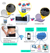
FIGURE 1
FIGURE 1
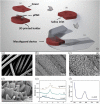
(i) Schematic diagram showing the fabrication process of Ni-Co-S@CF electrodes (Hekmat et al., 2021). (ii) Prospective toward the long-term glucose monitoring application of point-of-care wearable glucose sensors, iGLU 2.0 (Joshi et al., 2020).
FIGURE 2
FIGURE 2
(i) Schematic illustration demonstrating the fabrication of the self-powered sensor for point-of-care sweat monitoring (A), a photograph of the fabricated sensor (B), and photographs of the point-of-care sensor on the forehead of the volunteer when exercising 0 min (C), after 29-min exercise (D), and after 32-min exercise (E) (Zhang et al., 2018a). (ii) Working of the WCECS in real time on the body. (A) Photograph of the WCECS attached on the back of a human subject. (B) EC response of sweat glucose in the post-meal and fasting state. (C) Contrast of the sweat glucose concentrations sensed by the WCECS glucometer and glucose test kit. (D) Comparison of the glucose concentrations detected in 1 day by the glucometer, glucose test kit, and WCECS. (E) Evaluation of durability of the WCECS (Zheng et al., 2021).
FIGURE 3
FIGURE 3
(i) (A) Different skin conditions for artificial skin and the sensor tied on the human skin. (B) Equivalent circuit representation of the designed RuS2/PDMS-based hydration sensor. (C) Impedance value detected at the increase in humidity conditions on artificial skin at an alternating current frequency of 10 kHz. (D) Capacitance and resistance as the function of the increase in relative humidity conditions when the sensor was tied on the human skin. (E) Capacitance and resistance values of the human skin and artificial skin at distinct hydration environments (Veeralingam et al., 2020). (ii) (A) Immunoassay with the ability of the combined monitoring of glucose and alcohol. (B) Sweat sensor array displaying fluid confinement in the active detection region, size comparison with one cent, and the flexibility of sensor (Bhide et al., 2018a).
FIGURE 4
FIGURE 4
(A) Photograph of the wearable patch-based glucose sensor with a waterproof band and a sweat collection layer. (B) Photograph of wearable patch-based glucose sensor under deformation. (C) Optical image of disposable patch-based glucose sensor on the human skin with sweat (Lee et al., 2017).
FIGURE 5
FIGURE 5
Optical images and schematic diagrams displaying the wearable point-of-care biosensor toward the detection of glucose in perspiration. Photographs (A,B) of the constructed wearable sensor. Schematic illustration of the whole wearable point-of-care sensor (C) and exploded view (D) (Xuan et al., 2018).
FIGURE 6
FIGURE 6
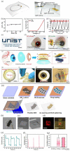
(i) Schematic illustration indicating the μPAD assembled into a mouth guard by a 3D-printed holder to form the wearable paper-based devices for point-of-care testing of glucose concentration in saliva. (A), (B), and (C) illustrate the arrangement of the μPAD in the 3D-printed holder, the final device before and after integration into the mouth guard, respectively (de Castro et al., 2019). (ii) Photographs (A) of the button-sensor, and schematic illustration showing (B) the assay procedure (Wei et al., 2021).
FIGURE 7
FIGURE 7
(i) Wireless representation circuit on the substrate. (A) Schematic diagram illustrating the wireless display circuit. The stretchable, transparent AgNF-based antenna and interconnects are in an elastic area, while the LED and rectifier are located in the reinforced area. (B) Relative change in transmitted voltage by antenna versus the applied strain. (C) Characterizations of Si diode on the hybrid substrate by using 0 and 30% in tensile strain. (D) Rectified properties of the constructed rectifier. (E) Optical image of wireless display circuit on the hybrid substrate. Scale bar, 1 cm. (F) Photos (left, off-state; right, on-state) of operating wireless display with lens shape located on the artificial eye. Scale bars, 1 cm (Ruan et al., 2017). (ii) Design of the structure of a smart contact lens with ultrathin MoS2 transistor–based serpentine mesh sensor system. (A) Schematic diagram showing the distinct layers of smart contact lens structure placed onto an eyeball. The dashed region highlights the method of gold-mediated mechanical exfoliation for the fabrication of monolayer MoS2. (B) Images of the sensor structure and serpentine electrode. (C) Photograph of a dome-shaped PDMS substrate with the sensor layer on it. (D) Photograph of an artificial eye with the sensing system attached to it. (E) Schematic diagram illustrating the smart contact lens and the sensors placed on the eyeball (Guo et al., 2021). (iii) On-demand drug delivery applying an f-DDS. (A) Schematic diagram displaying the construction process of f-DDS. (B) Photographic image of f-DDS. (C) SEM images of f-DDS before and after the gold electrochemistry experiment. Scale bar, 250 μm. (D) Confocal fluorescence microscopic images of rhodamine B dye released from drug reservoirs. Scale bars, 300 μm (left) and 500 μm (right). (E) Change of current for the f-DDS. (F) Released levels of genistein in a pulsatile manner. (G) Normalized content of genistein released from the reservoirs (n = 6) in comparison with the initial loading content (Keum et al., 2020).
FIGURE 8
FIGURE 8
Schematic illustration of the operation of the point-of-care device (Nightingale et al., 2019).
FIGURE 9
FIGURE 9
Schematic diagram illustrating the circuit illustration of the alarm glucose monitoring system and components of the wearable urine glucose biosensor system (Zhang et al., 2021a).

Similar articles

Cited by

References

    1. Adeel M., Rahman M. M., Caligiuri I., Canzonieri V., Rizzolio F., Daniele S. (2020). Recent Advances of Electrochemical and Optical Enzyme-free Glucose Sensors Operating at Physiological Conditions. Biosens. Bioelectron. 165, 112331. 10.1016/j.bios.2020.112331 - DOI - PubMed
    1. Arakawa T., Kuroki Y., Nitta H., Chouhan P., Toma K., Sawada S.-I., et al. (2016). Mouthguard Biosensor with Telemetry System for Monitoring of Saliva Glucose: A Novel Cavitas Sensor. Biosens. Bioelectron. 84, 106–111. 10.1016/j.bios.2015.12.014 - DOI - PubMed
    1. American Diabetes Association (2014). Diagnosis and Classification of Diabetes Mellitus. Diabetes Care 37, S81. 10.2337/dc14-S081 - DOI - PubMed
    1. Bandodkar A. J., Jia W., Yardımcı C., Wang X., Ramirez J., Wang J. (2015). Tattoo-based Noninvasive Glucose Monitoring: a Proof-Of-Concept Study. Anal. Chem. 87 (1), 394–398. 10.1021/ac504300n - DOI - PubMed
    1. Bandodkar A. J., Wang J. (2014). Non-invasive Wearable Electrochemical Sensors: a Review. Trends Biotechnol. 32 (7), 363–371. 10.1016/j.tibtech.2014.04.005 - DOI - PubMed

LinkOut - more resources