

Isolation and Characterization of Highly Pure Type A Spermatogonia From Sterlet (Acipenser ruthenus) Using Flow-Cytometric Cell Sorting
- PMID: 34957105
- PMCID: PMC8708567
- DOI: 10.3389/fcell.2021.772625
Isolation and Characterization of Highly Pure Type A Spermatogonia From Sterlet (Acipenser ruthenus) Using Flow-Cytometric Cell Sorting
Abstract
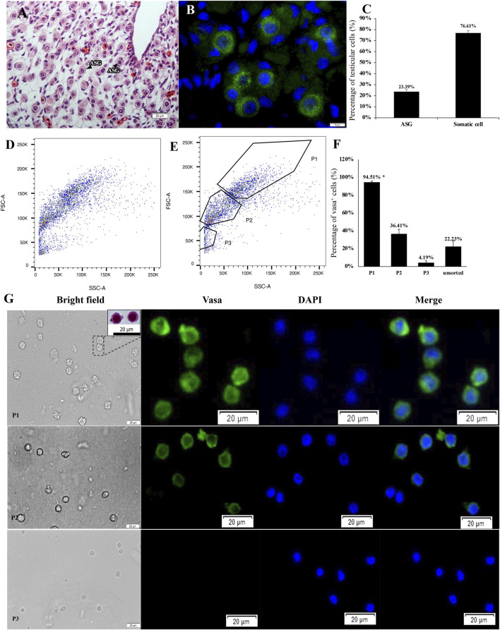
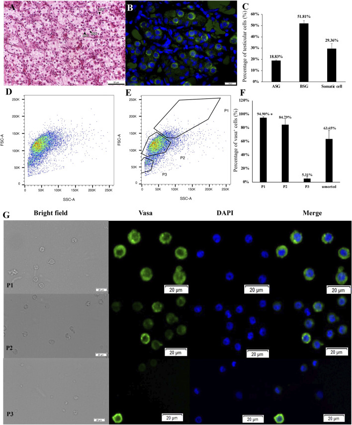
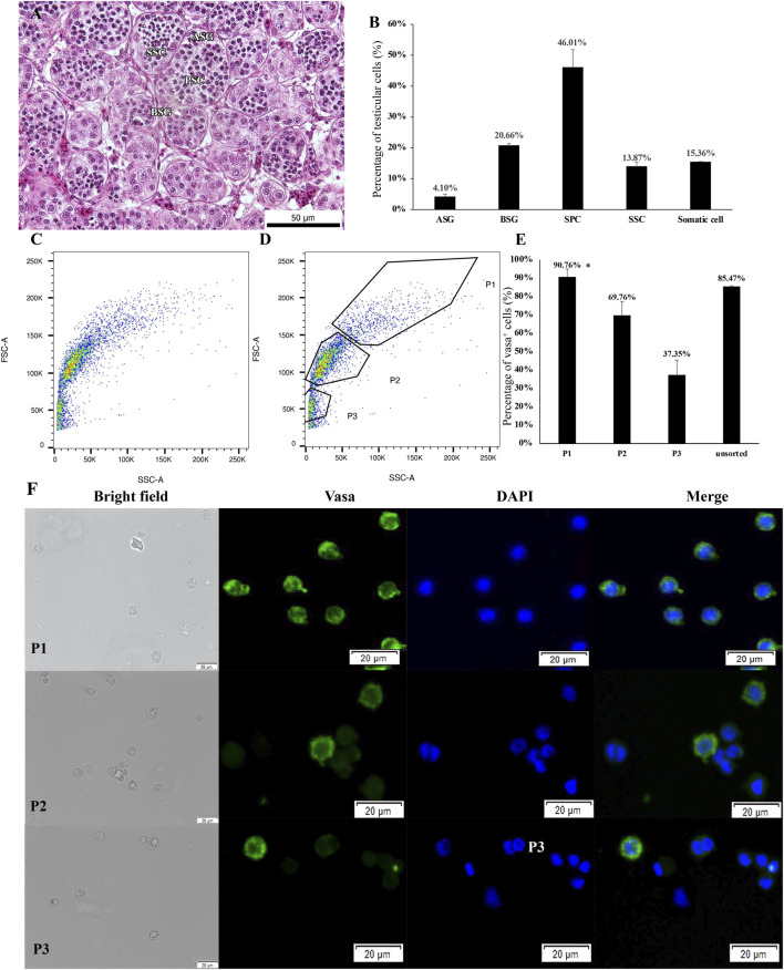
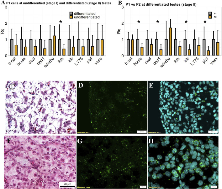
Sturgeons are among the most ancient linages of actinopterygians. At present, many sturgeon species are critically endangered. Surrogate production could be used as an affordable and a time-efficient method for endangered sturgeons. Our study established a method for identifying and isolating type A spermatogonia from different developmental stages of testes using flow cytometric cell sorting (FCM). Flow cytometric analysis of a whole testicular cell suspension showed several well-distinguished cell populations formed according to different values of light scatter parameters. FCM of these different cell populations was performed directly on glass slides for further immunocytochemistry to identify germ cells. Results showed that the cell population in gate P1 on a flow cytometry plot (with high forward scatter and high side scatter parameter values) contains the highest amount of type A spermatogonia. The sorted cell populations were characterized by expression profiles of 10 germ cell specific genes. The result confirmed that setting up for the P1 gate could precisely sort type A spermatogonia in all tested testicular developmental stages. The P2 gate, which was with lower forward scatter and side scatter values mostly, contained type B spermatogonia at a later maturing stage. Moreover, expressions of plzf, dnd, boule, and kitr were significantly higher in type A spermatogonia than in later developed germ cells. In addition, plzf was firstly found as a reliable marker to identify type A spermatogonia, which filled the gap of identification of spermatogonial stem cells in sterlet. It is expected to increase the efficiency of germ stem cell culture and transplantation with plzf identification. Our study thus first addressed a phenotypic characterization of a pure type A spermatogonia population in sterlet. FCM strategy can improve the production of sturgeons with surrogate broodstock and further the analysis of the cellular and molecular mechanisms of sturgeon germ cell development.
Keywords: PLZF; fluorescence-activated cell sorting; germ stem cell; gonad; spermatogonia; sturgeon.
Copyright © 2021 Xie, Tichopád, Kislik, Langerová, Abaffy, Šindelka, Franěk, Fučíková, Steinbach, Shah, Šauman, Chen and Pšenička.
Conflict of interest statement
The authors declare that the research was conducted in the absence of any commercial or financial relationships that could be construed as a potential conflict of interest.
Figures
Similar articles
-
Sterilization of sterlet Acipenser ruthenus by using knockdown agent, antisense morpholino oligonucleotide, against dead end gene.Theriogenology. 2015 Oct 15;84(7):1246-1255.e1. doi: 10.1016/j.theriogenology.2015.07.003. Epub 2015 Jul 11. Theriogenology. 2015. PMID: 26248520
-
Flow-cytometric enrichment of Pacific bluefin tuna type A spermatogonia based on light-scattering properties.Theriogenology. 2017 Oct 1;101:91-98. doi: 10.1016/j.theriogenology.2017.06.022. Epub 2017 Jun 22. Theriogenology. 2017. PMID: 28708521 Review.
-
Stage-specific embryonic antigen 4 is a membrane marker for enrichment of porcine spermatogonial stem cells.Andrology. 2020 Nov;8(6):1923-1934. doi: 10.1111/andr.12870. Epub 2020 Aug 9. Andrology. 2020. PMID: 32691968
-
Flow-cytometric isolation and enrichment of teleost type A spermatogonia based on light-scattering properties.Biol Reprod. 2012 Apr 12;86(4):107. doi: 10.1095/biolreprod.111.093161. Print 2012 Apr. Biol Reprod. 2012. PMID: 22219211
-
Spermatogonial stem cells: updates from specification to clinical relevance.Hum Reprod Update. 2019 May 1;25(3):275-297. doi: 10.1093/humupd/dmz006. Hum Reprod Update. 2019. PMID: 30810745 Review.
Cited by
-
Spermatogonial stem cell technologies: applications from human medicine to wildlife conservation†.Biol Reprod. 2024 Oct 14;111(4):757-779. doi: 10.1093/biolre/ioae109. Biol Reprod. 2024. PMID: 38993049 Free PMC article. Review.
-
Advantages, Factors, Obstacles, Potential Solutions, and Recent Advances of Fish Germ Cell Transplantation for Aquaculture-A Practical Review.Animals (Basel). 2022 Feb 10;12(4):423. doi: 10.3390/ani12040423. Animals (Basel). 2022. PMID: 35203131 Free PMC article. Review.
References
-
- Bemis W. E., Findeis E. K., Grande L. (1997). An Overview of Acipenseriformes. Environ. Biol. Fishes 48, 25–71. 10.1023/A:1007370213924 - DOI
-
- Bemis W. E., Kynard B. (1997). Sturgeon Rivers: an Introduction to Acipenseriform Biogeography and Life History. Environ. Biol. Fishes 48, 167–183. 10.1023/a:1007312524792 - DOI
LinkOut - more resources
Full Text Sources
Miscellaneous