. 2021 Dec 9:8:777685.
doi: 10.3389/fmed.2021.777685. eCollection 2021.

Agreement and Repeatability of Central and Peripheral Refraction by One Novel Multispectral-Based Refractor

Affiliations

Agreement and Repeatability of Central and Peripheral Refraction by One Novel Multispectral-Based Refractor

Weicong Lu et al. Front Med (Lausanne). .

Abstract

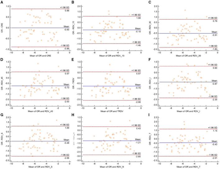
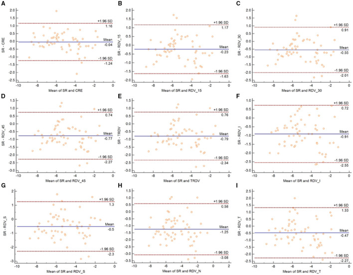
Purpose: To evaluate the repeatability of a multispectral-based refractor in central and peripheral refraction measurement, and to assess the agreement of such measurements with objective refraction (OR) and subjective refraction (SR) in patients with myopia. Methods: A total of 60 subjects were recruited in this prospective research. Patients were divided into three groups according to the refractive error. Next, the central and peripheral refraction parameters were measured using multispectral refractive tomography (MRT) before and after cycloplegia. In addition, OR and SR measurements were also performed. The intraobserver repeatability was analyzed using within-subject standard deviation (Sw), test-retest repeatability (TRT), and intraclass correlation coefficient (ICC). Agreement was evaluated using Bland-Altman plot and 95% limits of agreement (LoA). Results: The ICC value of central and peripheral refraction were all higher than 0.97 with or without cycloplegia. The peripheral refraction in the nasal, temporal, superior, and inferior quadrants was slightly worse than other parameters, with the largest error interval being 1.43 D. The 95% LoA of the central refraction and OR or SR ranged from -0.89 to 0.88 D and -1.24 to 1.16 D without cycloplegia, respectively, and from -0.80 to 0.42 D and -1.39 to -0.84 D under cycloplegia, respectively. Conclusions: The novel multispectral refraction topography demonstrated good repeatability in central and peripheral refraction. However, the refraction in the nasal, temporal, superior, and inferior quadrants were not as good as that of central and circle peripheral refraction.

Keywords: agreement; myopia; ophthalmology; refraction; repeatability.


Conflict of interest statement

The authors declare that the research was conducted in the absence of any commercial or financial relationships that could be construed as a potential conflict of interest.

Figures

Figure 1
Figure 1
MRT principle diagram. (A) Light ray radiation and sensor system. (B) Blurred image point with the change of focal length of fundus camera. (C) The sharpness profile of changing focal length.
Figure 2
Figure 2
The result provided by the MRT analysis system. (a) The relative peripheral refraction defocus value. (b) The block-refraction of absolute refraction value. (c,d) A direct view of the relative fraction status of the retinal by three-dimensional images viewing from superotemporal and inferonasal, respectively.
Figure 3
Figure 3
Bland–Altman plots between SR and CRE (A), RDV-15 (B), RDV-30 (C), RDV-45 (D), TRVD (E), RDV-I (F), RDV-S (G), RDV-N (H), and RDV-T (I) in non-cycloplegia eyes. The solid line shows the mean difference (bias), and the upper and lower lines represent 95% limits of agreement.
Figure 4
Figure 4
Bland–Altman plots between OR and CRE (A), RDV-15 (B), RDV-30 (C), RDV-45 (D), TRVD I (E), RDV-I (F), RDV-S (G), RDV-N (H), and RDV-T (I) in non-cycloplegia eyes. The solid line shows the mean difference (bias), and the upper and lower lines represent 95% limits of agreement.
Figure 5
Figure 5
Bland–Altman plots between SR and CRE (A), RDV-15 (B), RDV-30 (C), RDV-45 (D), TRVD (E), RDV-I (F), RDV-S (G), RDV-N (H), and RDV-T (I) in cycloplegia eyes. The solid line shows the mean difference (bias), and the upper and lower lines represent 95% limits of agreement.
Figure 6
Figure 6
Bland–Altman plots between OR and CRE (A), RDV-15 (B), RDV-30 (C), RDV-45 (D), TRVD (E), RDV-I (F), RDV-S (G), RDV-N (H), and RDV-T (I) in cycloplegia eyes. The solid line shows the mean difference (bias), and the upper and lower lines represent 95% limits of agreement.

References

    1. Huang J, Wen D, Wang Q, McAlinden C, Flitcroft I, Chen H, et al. . Efficacy comparison of 16 interventions for myopia control in children: a network meta-analysis. Ophthalmology. (2016) 123:697–708. 10.1016/j.ophtha.2015.11.010 - DOI - PubMed
    1. Baird PN, Saw SM, Lanca C, Guggenheim JA, Smith EL, Iii, Zhou X, et al. . Myopia. Nat Rev Dis Primers. (2020) 6:99. 10.1038/s41572-020-00231-4 - DOI - PubMed
    1. Pan CW, Ramamurthy D, Saw SM. Worldwide prevalence and risk factors for myopia. Ophthal Physiol Opt. (2012) 32:3–16. 10.1111/j.1475-1313.2011.00884.x - DOI - PubMed
    1. Morgan I, Rose K. How genetic is school myopia? Prog Retinal Eye Res. (2005) 24:1–38. 10.1016/j.preteyeres.2004.06.004 - DOI - PubMed
    1. Smith EL, Hung LF, Huang J. Relative peripheral hyperopic defocus alters central refractive development in infant monkeys. Vis Res. (2009) 49:2386–92. 10.1016/j.visres.2009.07.011 - DOI - PMC - PubMed