. 2022 Oct 21;145(10):3582-3593.
doi: 10.1093/brain/awab478.

APOE4 derived from astrocytes leads to blood-brain barrier impairment

Affiliations

APOE4 derived from astrocytes leads to blood-brain barrier impairment

Rosemary J Jackson et al. Brain. .

Abstract

Apolipoprotein E (ApoE) is a multifaceted secreted molecule synthesized in the CNS by astrocytes and microglia, and in the periphery largely by the liver. ApoE has been shown to impact the integrity of the blood-brain barrier, and, in humans, the APOE4 allele of the gene is reported to lead to a leaky blood-brain barrier. We used allele specific knock-in mice expressing each of the common (human) ApoE alleles, and longitudinal multiphoton intravital microscopy, to directly monitor the impact of various ApoE isoforms on blood-brain barrier integrity. We found that humanized APOE4, but not APOE2 or APOE3, mice show a leaky blood-brain barrier, increased MMP9, impaired tight junctions, and reduced astrocyte end-foot coverage of blood vessels. Removal of astrocyte-produced ApoE4 led to the amelioration of all phenotypes while the removal of astrocyte-produced ApoE3 had no effect on blood-brain barrier integrity. This work shows a cell specific gain of function effect of ApoE4 in the dysfunction of the BBB and implicates astrocyte production of ApoE4, possibly as a function of astrocytic end foot interactions with vessels, as a key regulator of the integrity of the blood-brain barrier.

Keywords: Alzheimer's disease; apolipoprotein E; astrocytes; blood–brain barrier.


Figures

Figure 1
Figure 1
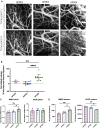
ApoE4-TR mice have an impaired BBB compared with ApoE3-TR and ApoE2-TR mice. (A) Multiphoton microscopy of 40 kDa fluorescein conjugated dextran in 9-month-old TR-APOE2, TR-APOE3 and TR-APOE4 mice. Images are a z-projection of ∼120 μm of cortex. (B) Quantification of leakage ANOVA [F(2,27) = 25.03, P < 0.0001]. (C) Analysis of whole brain mRNA levels show an increase in MMP9 in APOE4 animals ANOVA [F(2,14) = 9.086, P = 0.0030] and APOE mRNA levels are unchanged between the three APOE genotypes. (D) Analysis of whole brain protein levels of MMP9 show an increase in MMP9 in APOE4 animals compared with APOE2 and APOE3 animals one-way ANOVA [F(2,9) = 10.01, P = 0.0052] and a decrease in APOE protein levels in the APOE4 animals as compared with the APOE2 and APOE3 animals APOE ANOVA [F(2,9) = 12.84, P = 0.0023]. Post hoc test results are represented as stars *P < 0.05, **P < 0.01, ***P < 0.001, ****P < 0.0001.
Figure 2
Figure 2
Tamoxifen injection induces a lasting knockout of APOE from astrocytes specifically. (A) PCR of the flox stop site indicates that CRE recombination has occurred in animals that have the ALDH1L1 CRE driver and have been injected with tamoxifen. No recombination event was observed in mice lacking the driver (ALDH1L1-Cre−) or injected with corn oil vehicle only (Tam−). (B) APOE qPCR shows a 40–50% decrease in APOE mRNA in the brain parenchyma after knocking down APOE expressing in astrocytes compared with mice expressing APOE in all cells ANOVA [F(3,34) = 16.90, P < 0.0001]. (C) RNAscope shows knockout of APOE mRNA in GFAP astrocytes in the CNS. (D) Western blot shows a 40–50% knock down of APOE protein in astrocyte knockout mice ANOVA [F(3,35) = 121.05, P < 0.0001. (E) Western blot shows that APOE4 mice show no effect of tamoxifen injection on APOE levels and that APOE aKO mice show no effect 1 week following tamoxifen injection but that by 1 month APOE protein levels are reduced by 40–50% and this is sustained at 2 months ANOVA [F(6,25) = 49.36, P < 0.0001. (F) Western blot shows a 15% knock down of plasma APOE protein in APOE3 aKO and APOE4aKO mice ANOVA [F(3,13) = 9.571, P = 0.0013].
Figure 3
Figure 3
Astrocyte-specific knockout of APOE4 rescues BBB impairments. (A) Timeline of in vivo studies. (B) Multiphoton microscopy or 40 kDa fluorescein conjugated dextran in ALDH1L1CRE × TR-APOE4 mice before administration of tamoxifen and 2 months post-tamoxifen injection. Images are a z-projection of 100 μm of cortex. (C) Quantification of BBB leakage over time in ALDH1L1CRE × TR-APOE4 compared with 9-month TR-APOE3 mice shows a significant leakage prior to and 1 week after tamoxifen injection. At 1 and 2 months after tamoxifen injection, leakage was rescued down to APOE3 levels ANOVA [F(4,32) = 36.01, P < 0.0001]. (D) Quantification of BBB leakage 2 months after tamoxifen injection for TR-APOE3, ALDH1L1Cre × TR-APOE3, TR-APOE4 and ALDH1L1Cre × TR-APOE4 shows that APOE4 mice show significant leakage but that APOE3, APOE3 aKO and APOE4 aKO mice do not ANOVA [F(3,16) = 36.01, P = 0.0003]. (E) Accumulation of fluorescence in the brain parenchyma over time in 12-month APOE3, APOE3 aKO APOE4, and APOE4 aKO mice shows that only APOE4 mice have a slope that is significantly different from 0 [F(1,26) = 11.57, P = 0.0022]. (F) We saw no differences in measured vessel width. Note, not all mice were imaged at all time points due to COVID-19 restrictions and thus n numbers fluctuated; however, each mouse is shown as an individual point on the graph (n numbers for E: APOE3 n = 6, APOE4 n = 6, APOE3 aKO n = 3, APOE4, aKO n = 5).
Figure 4
Figure 4
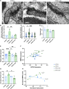
Tight junctions are impaired in ApoE4 mice along with MMP9 expression. (A and B) Electron microscopy analysis shows that APOE4 mice show an increased distance (as indicated by a small line) between membranes at tight junctions (white arrows) and that this is ameliorated in APOE4 astrocyte knockout mice both when compared at the mouse level F(3,11) = 5.926 P = 0.0117 and (C) the individual tight junction F(3,94) = 3.314 P = 0.0233. (D) However, we saw no different in the thickness of the endothelium. (E) Analysis of whole brain mRNA show that APOE4 animals have significantly higher expression of MMP9 compared with APOE3 animals but that knock of APOE4 from astrocytes reduces MMP9 expression to the level of APOE3 animals, APOE3 knockout has no effect F(3,32) = 8.153 P = 0.0004. (F) There is a correlation between APOE4 expression and MMP9 expression (P = 0.0021) but no correlation between these genes in APOE3 mice. (G) Analysis of protein whole brain by western blot shows that there is an increase in MMP9 protein in APOE4 animals compared with APOE3 animals and that astrocyte knock out reduces the MMP9 protein levels of APOE3 animals F(3,11) = 6.115 P = 0.0106. (H) There is a positive correlation at the protein level between APOE4 expression and MMP9 expression (P = 0.0018) and a negative correlation between these genes in APOE3 mice (P = 0.0418).
Figure 5
Figure 5
Astrocyte end-feet are impaired in APOE4 mice. (A) Analysis by electron microscopy shows that there is a decreased coverage of astrocytic end-feet (in yellow) in APOE4 mice when compared with APOE3 mice (B) and that this is ameliorated in the APOE4 astrocyte knockout mice. F(3,11) = 5.926 P = 0.0117. (C) Vessel perimeter did not differ between groups F(3,11) = 0.4780 P = 0.7040.

References

    1. Keaney J, Campbell M. The dynamic blood-brain barrier. FEBS J. 2015;282(21):4067–4079. - PubMed
    1. Sweeney MD, Sagare AP, Zlokovic BV. Blood-brain barrier breakdown in Alzheimer disease and other neurodegenerative disorders. Nat Rev Neurol. 2018;14(3):133–150. - PMC - PubMed
    1. Bowman GL, Dayon L, Kirkland R, et al. . Blood-brain barrier breakdown, neuroinflammation, and cognitive decline in older adults. Alzheimer’s Dement. 2018;14(12):1640–1650. - PubMed
    1. Cortes-Canteli M, Iadecola C. Alzheimer’s disease and vascular aging: JACC focus seminar. J Am Coll Cardiol. 2020;75(8):942–951. - PMC - PubMed
    1. Linton MF, Gish R, Hubl ST, et al. . Phenotypes of apolipoprotein B and apolipoprotein E after liver transplantation. J Clin Invest. 1991;88(1):270–281. - PMC - PubMed

Publication types