. 2022 Jan 19;33(1):194-205.
doi: 10.1021/acs.bioconjchem.1c00537. Epub 2021 Dec 25.

Strain-Promoted Azide-Alkyne Cycloaddition-Based PSMA-Targeting Ligands for Multimodal Intraoperative Tumor Detection of Prostate Cancer

Affiliations

Strain-Promoted Azide-Alkyne Cycloaddition-Based PSMA-Targeting Ligands for Multimodal Intraoperative Tumor Detection of Prostate Cancer

Yvonne H W Derks et al. Bioconjug Chem. .

Abstract

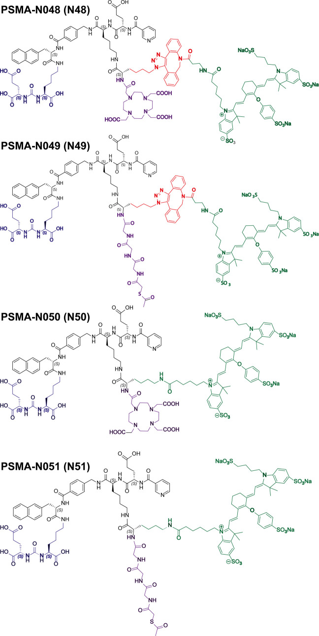
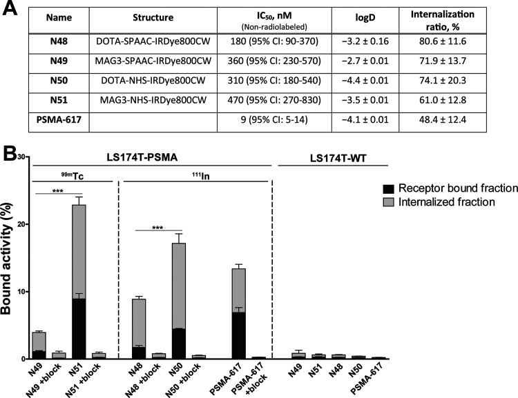
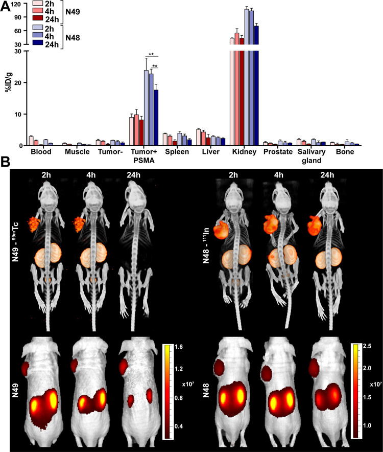
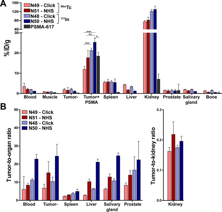
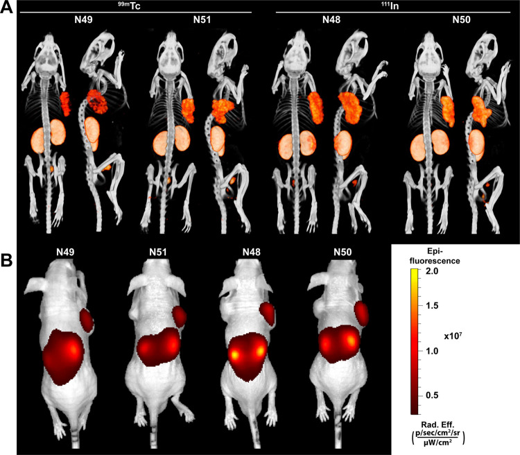
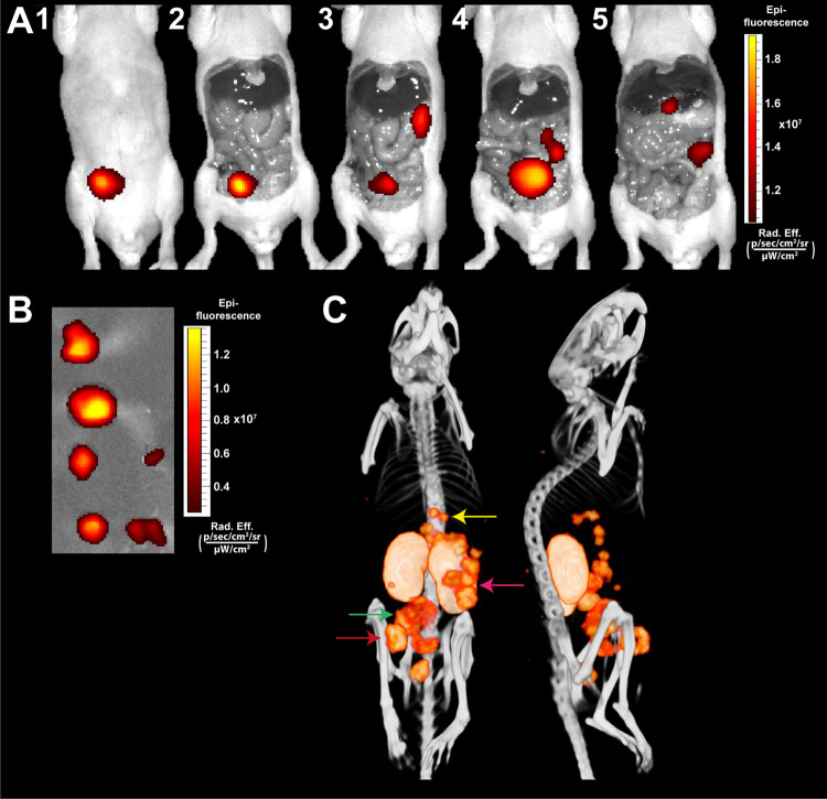
Strain-promoted azide-alkyne cycloaddition (SPAAC) is a straightforward and multipurpose conjugation strategy. The use of SPAAC to link different functional elements to prostate-specific membrane antigen (PSMA) ligands would facilitate the development of a modular platform for PSMA-targeted imaging and therapy of prostate cancer (PCa). As a first proof of concept for the SPAAC chemistry platform, we synthesized and characterized four dual-labeled PSMA ligands for intraoperative radiodetection and fluorescence imaging of PCa. Ligands were synthesized using solid-phase chemistry and contained a chelator for 111In or 99mTc labeling. The fluorophore IRDye800CW was conjugated using SPAAC chemistry or conventional N-hydroxysuccinimide (NHS)-ester coupling. Log D values were measured and PSMA specificity of these ligands was determined in LS174T-PSMA cells. Tumor targeting was evaluated in BALB/c nude mice with subcutaneous LS174T-PSMA and LS174T wild-type tumors using μSPECT/CT imaging, fluorescence imaging, and biodistribution studies. SPAAC chemistry increased the lipophilicity of the ligands (log D range: -2.4 to -4.4). In vivo, SPAAC chemistry ligands showed high and specific accumulation in s.c. LS174T-PSMA tumors up to 24 h after injection, enabling clear visualization using μSPECT/CT and fluorescence imaging. Overall, no significant differences between the SPAAC chemistry ligands and their NHS-based counterparts were found (2 h p.i., p > 0.05), while 111In-labeled ligands outperformed the 99mTc ligands. Here, we demonstrate that our newly developed SPAAC-based PSMA ligands show high PSMA-specific tumor targeting. The use of click chemistry in PSMA ligand development opens up the opportunity for fast, efficient, and versatile conjugations of multiple imaging moieties and/or drugs.


Conflict of interest statement

The authors declare the following competing financial interest(s): Y. Derks, D. Lwik, S. Heskamp, P. Laverman and M. Rijpkema are applicants of patent: EP21155853 - PSMA-targeting ligands for multimodal applications. No other potential conflicts of interest relevant to this article exist.

Figures

Figure 1
Figure 1
Chemical structures of PSMA-N048 (N48), PSMA-N049 (N49), PSMA-N050 (N50), and PSMA-N051 (N51). Ligands consisting of KuE (blue), linker (black), SPAAC (red), IRDye800CW (green), and MAG3 or DOTA chelator (purple).
Figure 2
Figure 2
In vitro characterization of N48, N49, N50, and N51. (A) IC50 values of ligands as determined in competitive binding assays using LS174T-PSMA cells. IC50 values were determined using a nonradiolabeled ligand (N48, N49, N50, and N51) in competition with 111In-labeled PSMA-617. Lipophilicity of ligands expressed in log D values. Internalization ratio as determined in LS174T-PSMA cells. (B) Membrane binding and internalization kinetics of N48, N49, N50, and N51 in LS174T-PSMA-positive and -negative cells. Nonspecific binding was determined by blocking with an excess of 2-PMPA (50 μg). PSMA-617 was added as a positive control.
Figure 3
Figure 3
In vivo pharmacokinetics of 99mTc-N49 and 111In-N48. (A) Biodistribution as determined after dissection of 99mTc-N49 (3 MBq/mouse) and 111In-N48 (10 MBq/mouse) 2, 4, and 24 h p.i. (0.3 nmol, n = 5/group). Biodistribution was determined in mice bearing subcutaneous LS174T-PSMA and LS174T wild-type xenografts. Data are expressed as %ID/g ± SD; ** indicates p < 0.01. (B) Representative μSPECT/CT scans and fluorescence images of mice with s.c. LS174T-PSMA (left) and wild-type LS174T (right) tumors after i.v. injection of 99mTc-N49 (3 MBq/mouse) and 111In-N48 (10 MBq/mouse) 2, 4, and 24 h p.i.
Figure 4
Figure 4
In vivo comparison of N48, N49, N50, and N51. (A) Biodistribution as determined after dissection and (B) resulting tumor-to-organ ratios of four 111In- (10 MBq/mouse) or 99mTc-labeled (15 MBq/mouse) ligands and positive control PSMA-617 (0.3 nmol, 2 h p.i., n = 5/group). Biodistribution was determined in mice bearing subcutaneous LS174T-PSMA and LS174T wild-type xenografts. Data are expressed as %ID/g ± SD; * indicates p < 0.05, ** indicates p < 0.01, and *** indicates p < 0.001.
Figure 5
Figure 5
All ligands clearly visualize PSMA-positive tumors using both μSPECT/CT and fluorescence imaging. Representative μSPECT/CT scans (A) and fluorescence images (B) of mice with s.c. LS174T-PSMA (right) and wild-type LS174T (left) tumors after i.v. injection of 111In- (10 MBq/mouse) or 99mTc-labeled (15 MBq/mouse) ligands (0.3 nmol, 2 h p.i.).
Figure 6
Figure 6
Multimodal fluorescence and μSPECT/CT imaging of intraperitoneal PSMA-positive tumors using 111In-N50. Mouse with several intraperitoneal LS174T-PSMA tumors located at different depths in the peritoneal cavity. (A) Same-scale NIRF images of mouse with several intraperitoneal tumors after i.v. injection of 111In-labeled N50 (0.3 nmol, 10 MBq/mouse, 2 h p.i.). (B) NIRF image of removed tumors. (C) Corresponding μSPECT/CT images in supine and left lateral positions.

References

    1. Sung H.; Ferlay J.; Siegel R. L.; Laversanne M.; Soerjomataram I.; Jemal A.; Bray F. Global cancer statistics 2020: GLOBOCAN estimates of incidence and mortality worldwide for 36 cancers in 185 countries. Ca-Cancer J. Clin. 2021, 71, 209.10.3322/caac.21660. - DOI - PubMed
    1. Wang X.; Huang S. S.; Heston W. D.; Guo H.; Wang B. C.; Basilion J. P. Development of targeted near-infrared imaging agents for prostate cancer. Mol. Cancer Ther. 2014, 13, 2595–2606. 10.1158/1535-7163.Mct-14-0422. - DOI - PMC - PubMed
    1. Lütje S.; Heskamp S.; Franssen G. M.; Frielink C.; Kip A.; Hekman M.; Fracasso G.; Colombatti M.; Herrmann K.; Boerman O. C.; et al. Development and characterization of a theranostic multimodal anti-PSMA targeting agent for imaging, surgical guidance, and targeted photodynamic therapy of PSMA-expressing tumors. Theranostics 2019, 9, 2924–2938. 10.7150/thno.35274. - DOI - PMC - PubMed
    1. Derks Y. H. W.; Löwik D. W. P. M.; Sedelaar J. P. M.; Gotthardt M.; Boerman O. C.; Rijpkema M.; Lütje S.; Heskamp S. PSMA-targeting agents for radio- and fluorescence-guided prostate cancer surgery. Theranostics 2019, 9, 6824–6839. 10.7150/thno.36739. - DOI - PMC - PubMed
    1. Lütje S.; Rijpkema M.; Franssen G. M.; Fracasso G.; Helfrich W.; Eek A.; Oyen W. J.; Colombatti M.; Boerman O. C. Dual-Modality Image-Guided Surgery of Prostate Cancer with a Radiolabeled Fluorescent Anti-PSMA Monoclonal Antibody. J. Nucl. Med. 2014, 55, 995–1001. 10.2967/jnumed.114.138180. - DOI - PubMed