. 2022;85(4):1555-1571.
doi: 10.3233/JAD-215025.

Probiotic Bifidobacterium breve Prevents Memory Impairment Through the Reduction of Both Amyloid-β Production and Microglia Activation in APP Knock-In Mouse

Affiliations

Probiotic Bifidobacterium breve Prevents Memory Impairment Through the Reduction of Both Amyloid-β Production and Microglia Activation in APP Knock-In Mouse

Mona Abdelhamid et al. J Alzheimers Dis. 2022.

Erratum in

Abstract

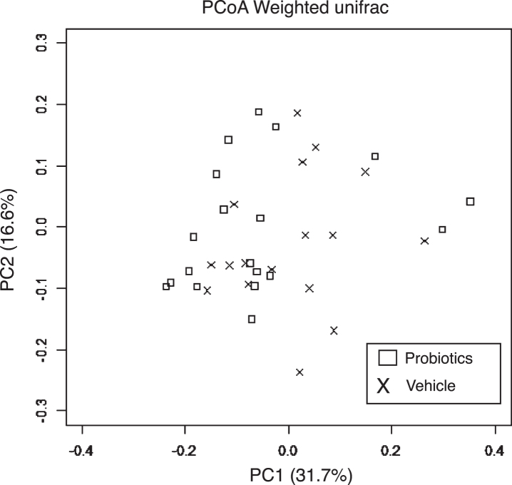
Background: Probiotic supplementation reestablishes microbiome diversity and improves brain function in Alzheimer's disease (AD); their molecular mechanisms, however, have not yet been fully illustrated.

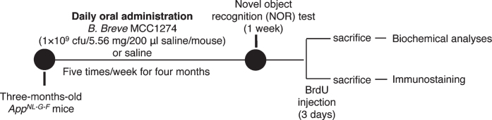
Objective: We investigated the effects of orally supplemented Bifidobacterium breve MCC1274 on cognitive function and AD-like pathologies in AppNL-G-F mice.

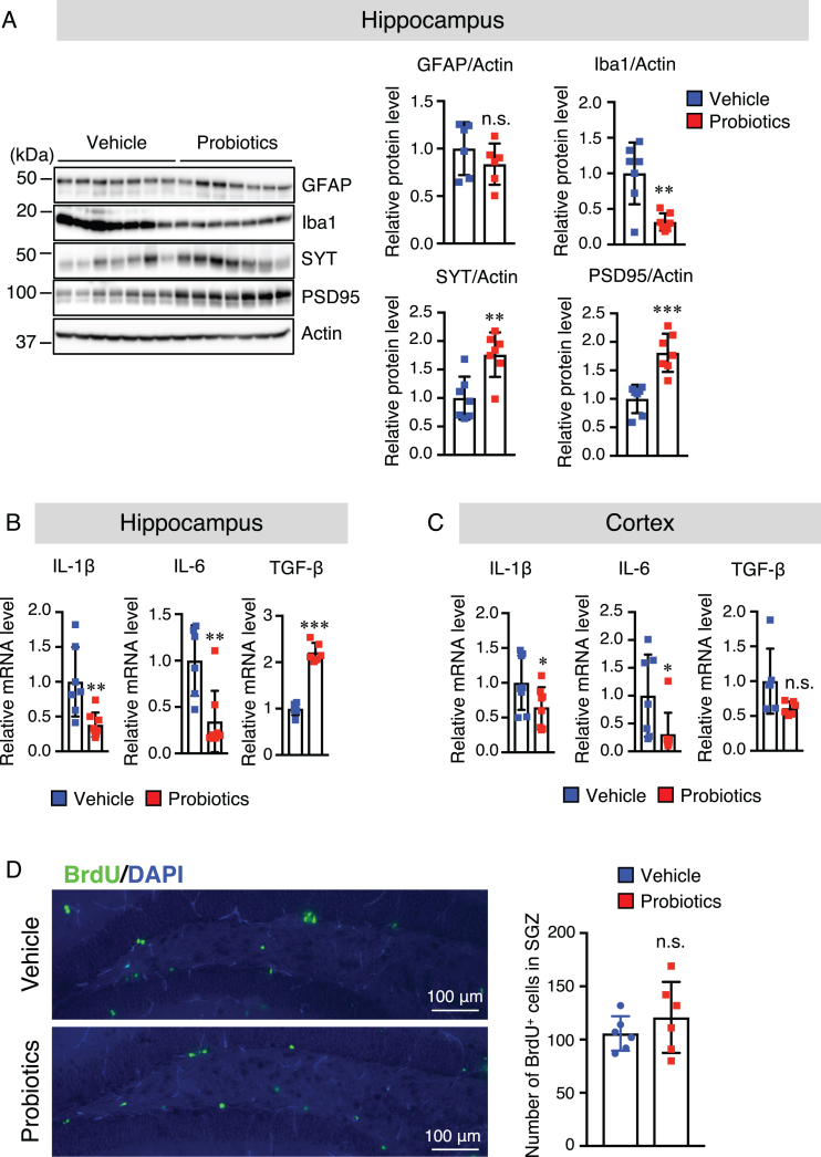
Methods: Three-month-old AppNL-G-F mice were orally supplemented with B. breve MCC1274 for four months. The short-term memory function was evaluated using a novel object recognition test. Amyloid plaques, amyloid-β (Aβ) levels, Aβ fibril, amyloid-β protein precursor and its processing enzymes, its metabolic products, glial activity, and cell proliferation in the subgranular zone of the dentate gyrus were evaluated by immunohistochemistry, Aβ ELISA, western blotting, and immunofluorescence staining. The mRNA expression levels of pro- and anti-inflammatory cytokines were determined by qRT-PCR analysis.

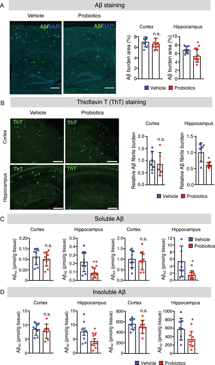
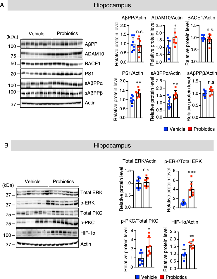
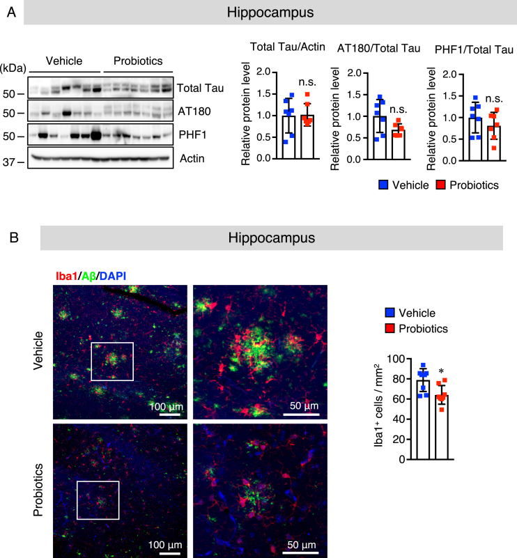
Results: We found that the oral B. breve MCC1 274 supplementation prevented memory impairment in AppNL-G-F mice and decreased hippocampal Aβ levels through the enhancement of the a-disintegrin and metalloproteinase 10 (ADAM10) level. Moreover, administration of the probiotic activated the ERK/HIF-1α signaling pathway responsible for increasing the ADAM10 level and also attenuated microglial activation, which in turn led to reduction in the mRNA expression levels of pro-inflammatory cytokines in the brain. In addition, B. breve MCC1274 supplementation increased the level of synaptic proteins in the hippocampus.

Conclusion: Our findings support the possibility that oral B. breve MCC1274 supplementation might be used as a potential preventive therapy for AD progression.

Keywords: ADAM10; Alzheimer’s disease; Bifidobacterium breve MCC1274; ERK; HIF-1α; amyloid-β production; glial activation; novel object recognition; synapses.


Conflict of interest statement

Authors’ disclosures available online (https://www.j-alz.com/manuscript-disclosures/21-5025r2).

Figures

Fig. 1
Fig. 1
Experimental procedure. Three-month-old AppNL-G-F mice were assigned randomly into the vehicle and probiotic groups: the vehicle group (n = 26) received saline and the probiotic group (n = 26) was supplemented with B. breve MCC1274 (1×10⁹ cfu/5.56 mg/200μl saline/mouse) via oral gavage five times/week for four months. At the end of supplementation, feces were collected, and then the mice were behaviorally evaluated by novel object recognition (NOR) test or injected with BrdU. The mice were sacrificed and brains collected for biochemical analyses and immunostaining.
Fig. 2
Fig. 2
B. breve MCC1274 supplementation reduces Aβ plaque burden, Aβ levels, and Aβ fibrils in the hippocampus of AppNL-G-F mice. A) The representative fluorescent images of Aβ plaque burden detected by anti-Aβ antibody (82E1), which recognizes both Aβ40 and Aβ42 (left panel). Aβ burden areas including the cortex and hippocampus were quantified as the percentage of immunostained area divided by all cortical and hippocampal areas (right panel). Scale bars are 250μm. B) The representative fluorescent images of Aβ fibril were detected by thioflavin T (left panel). Relative Aβ fibril burden in both cortex and hippocampus were quantified (right panel), Scale bars are 100μm. Sandwich ELISA result of cortical and hippocampal levels of soluble Aβ40 and Aβ42 (C) and insoluble Aβ40 and Aβ42 (D) of AppNL-G-F mice. Aβ levels were normalized to each tissue weight. Data are expressed as the mean±SD, n = 9-10, *p < 0.05, **p < 0.01 compared with the vehicle group, as determined by Student’s t-test.
Fig. 3
Fig. 3
B. breve MCC1274 supplementation upregulates ADAM10 protein level in the hippocampus of AppNL-G-F mice. Protein levels of AβPP, ADAM10, BACE1, PS1, sAβPPα, sAβPPβ, and actin (A) and protein levels of total ERK, phosphorylated (p) ERK, total PKC, pPKC, HIF-1α, and actin (B) in the hippocampus were determined by western blot analysis, quantified by densitometry, normalized to actin level, and expressed as a value relative to the control. Data are expressed as the mean±SD, n = 7. *p < 0.05, **p < 0.01, ***p < 0.001 compared with the vehicle group, n.s., no significant difference, as determined by Student’s t-test.
Fig. 4
Fig. 4
B. breve MCC1274 supplementation does not affect the phosphorylation of tau and attenuates microglial activation in the hippocampus of AppNL-G-F mice. A) The protein levels of total Tau, AT180 (p-Thr23), PHF1 (p-Ser369/Ser404), and actin in the hippocampus were determined by western blot analysis, quantified by densitometry, normalized to actin level, and expressed as a value relative to the control. B) Brain sections were stained with the anti-Iba1 (red) and anti-Aβ (green) antibodies, and cell nuclei were stained with DAPI (blue). Representative images of the hippocampus (left panel). Highly magnified images of the squared region in the left panels are shown in the adjacent right panels. Numbers of Iba1-positive cells (right panel) in the hippocampus. Data are expressed as the mean±SD, n = 6-7, *p < 0.05 compared with the vehicle group, n.s., no significant difference as determined by Student’s t-test.
Fig. 5
Fig. 5
B. breve MCC1274 supplementation decreases the Iba1 protein level and increases synaptic protein levels in the hippocampus, whereas it has no effect on cell proliferation in the DG of the hippocampus. A) Protein levels of GFAP, Iba1, SYT, PSD95, and actin in the hippocampus were determined by western blot analysis and quantified by densitometry, normalized to actin level, and expressed as a value relative to the control. IL-6 and IL-1β, and TFG-β1 mRNA expression levels in the hippocampus (B) and cortex (C) were assessed by qRT-PCR analysis. Each mRNA expression was normalized to the corresponding amount of GAPDH mRNA. D) Brain sections were stained with the anti-BrdU antibody (green) and cell nuclei were stained with DAPI (blue) in DG in the hippocampus (left panel). The number of BrdU+ cells in the SGZ of DG (right panel). Data are expressed as the mean±SD, n = 6-7, *p < 0.05, **p < 0.01, ***p < 0.001 compared with the vehicle group n.s., no significant difference as determined by Student’s t-test.
Fig. 6
Fig. 6
Effects of B. breve MCC1274 supplementation on gut microbiota. PCoA based on weighted unifrac distance from 16S rRNA sequencing data did not show a significant difference between AppNL-G-F mice with and without B. breve MCC1274 supplementation as determined by permutation MANOVA (p =0.379).

References

    1. Mount C, Downton C (2006) Alzheimer disease: progress or profit? Nat Med 12, 780–784. - PubMed
    1. Takasugi N, Tomita T, Hayashi I, Tsuruoka M, Niimura M, Takahashi Y, Thinakaran G, Iwatsubo T (2003) The role of presenilin cofactors in the gamma-secretase complex. Nature 422, 438–441. - PubMed
    1. Lammich S, Kojro E, Postina R, Gilbert S, Pfeiffer R, Jasionowski M, Haass C, Fahrenholz F (1999) Constitutive and regulated alpha-secretase cleavage of Alzheimer’s amyloid precursor protein by a disintegrin metalloprotease. Proc Natl Acad Sci U S A 96, 3922–3927. - PMC - PubMed
    1. Castro-Gomez S, Binder J, Heneka MT (2019) [Neuroinflammation as motor of Alzheimer’s disease]. Nervenarzt 90, 898–906. - PubMed
    1. Heneka MT, Carson MJ, El Khoury J, Landreth GE, Brosseron F, Feinstein DL, Jacobs AH, Wyss-Coray T, Vitorica J, Ransohoff RM, Herrup K, Frautschy SA, Finsen B, Brown GC, Verkhratsky A, Yamanaka K, Koistinaho J, Latz E, Halle A, Petzold GC, Town T, Morgan D, Shinohara ML, Perry VH, Holmes C, Bazan NG, Brooks DJ, Hunot S, Joseph B, Deigendesch N, Garaschuk O, Boddeke E, Dinarello CA, Breitner JC, Cole GM, Golenbock DT, Kummer MP (2015) Neuroinflammation in Alzheimer’s disease. Lancet Neurol 14, 388–405. - PMC - PubMed

Publication types

Substances