Meta-Analysis
. 2022 Jun;48(4):e12790.
doi: 10.1111/nan.12790. Epub 2022 Mar 3.

Diagnostic accuracy of 1p/19q codeletion tests in oligodendroglioma: A comprehensive meta-analysis based on a Cochrane systematic review

Affiliations
Meta-Analysis

Diagnostic accuracy of 1p/19q codeletion tests in oligodendroglioma: A comprehensive meta-analysis based on a Cochrane systematic review

Sebastian Brandner et al. Neuropathol Appl Neurobiol. 2022 Jun.

Abstract

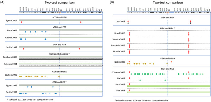
Codeletion of chromosomal arms 1p and 19q, in conjunction with a mutation in the isocitrate dehydrogenase 1 or 2 gene, is the molecular diagnostic criterion for oligodendroglioma, IDH mutant and 1p/19q codeleted. 1p/19q codeletion is a diagnostic marker and allows prognostication and prediction of the best drug response within IDH-mutant tumours. We performed a Cochrane review and simple economic analysis to establish the most sensitive, specific and cost-effective techniques for determining 1p/19q codeletion status. Fluorescent in situ hybridisation (FISH) and polymerase chain reaction (PCR)-based loss of heterozygosity (LOH) test methods were considered as reference standard. Most techniques (FISH, chromogenic in situ hybridisation [CISH], PCR, real-time PCR, multiplex ligation-dependent probe amplification [MLPA], single nucleotide polymorphism [SNP] array, comparative genomic hybridisation [CGH], array CGH, next-generation sequencing [NGS], mass spectrometry and NanoString) showed good sensitivity (few false negatives) for detection of 1p/19q codeletions in glioma, irrespective of whether FISH or PCR-based LOH was used as the reference standard. Both NGS and SNP array had a high specificity (fewer false positives) for 1p/19q codeletion when considered against FISH as the reference standard. Our findings suggest that G banding is not a suitable test for 1p/19q analysis. Within these limits, considering cost per diagnosis and using FISH as a reference, MLPA was marginally more cost-effective than other tests, although these economic analyses were limited by the range of available parameters, time horizon and data from multiple healthcare organisations.

Keywords: 1p/19q codeletion; PCR; false negative; false positive; fluorescent in situ hybridisation; oligodendroglioma.


Conflict of interest statement

The authors declare no conflicts of interest.

Figures

FIGURE 1
FIGURE 1
Graphical representation of absolute and relative 1p/19q codeletions. In all parts of the figure, chromosomes 1 and 19 are presented in separate frames to visualise the combination of FISH signals. The 1p and the 19q probes are red, and the reference probes (1q and 19p) are green. The approximate labelling sites are indicated in the chromosomal schematics. An unrelated chromosome (2) is also shown, and appearances as FISH images on the bottom of each frame. (A) Cell with diploid set of chromosomes, with two red signals each, for chromosomal arms 1p and 19q, as well as two green signals each for chromosomal arms 1q and 19p. (B) Absolute 1p/19q codeletion in a diploid set of chromosomes. Loss of one red signal in chromosome 1p and in 19q and two green signals for each 1q and 19p. (C) Relative codeletion with example of polysomy of chromosome 19 and chromosome 2, which has been suggested to indicate a worse prognosis [4, 5, 6, 7]. (D) 1p/19q codeletion in tetraploid cells, resulting in two red and four green signals for both 1p and 19q tests. (E) Complex deletion patterns as found in a small proportion of oligodendrogliomas, often associated with anaplastic histological types. In this example, there are diploid cells (left, 30%) triploid cells (centre, 30%) and tetraploid cells (right, 40%)
FIGURE 2
FIGURE 2
PRISMA flow chart illustrating the selection process of inclusions and exclusions of studies
FIGURE 3
FIGURE 3
Network plot of the included studies. The colour scheme of the circles corresponds to the colour scheme of the test methods represented in Figures 4, 5, 6, 7. The size of the circles represents the number of test results for a test category. The thickness of the lines is proportional to the number of studies making the comparison. Note that the FISH and PCR‐based LOH circles include within‐test category comparisons
FIGURE 4
FIGURE 4
(A) Graphical representation of regions analysed in studies comparing four tests: Blesa 2009 [30], Hatanpaa 2003 [31] and Duval 2014 [32], and (B) studies comparing three tests: Mohapatra 2006 [33], Pesenti 2017 [34], Burger 2001 [35], Smith 1999 [36], Dahlback 2011 [37], Belaud‐Rotureau 2006 [38], Horbinski 2012 [39] and Pesenti 2017 [34]. The top on both figures indicates a graphical representation of chromosome 1 (adapted from the GRCh38/hg38 Assembly). The figure legend indicates the different methods, with different colour codes for FISH, depending on the origin or manufacturer of the probes. In each section, the first author of the study is represented on top, and the techniques on the left of the table. All acronyms are explained in the main text
FIGURE 5
FIGURE 5
(A) Graphical representation of regions analysed in studies comparing two tests: aCGH and FISH (Byeon 2014 [40]), aCGH and PCR (Blesa 2009 [30] and Byeon 2014 [40]), CGH and FISH (Smith 1999 [36]), CGH and G banding (Dahlback 2009 [41] and Schrock 1994 [42]), CGH and MLPA (Jeuken 2006 [43]), and CGH and PCR (Bigner 1999 [44] and Smith 1999 [36]), and (B) CISH and FISH (Lass 2013 [45]), FISH and FISH (Duval 2015 [46], Senetta 2013 [47], Srebotnik‐Kirbis 2016 [48] and Uchida 2019 [49]), FISH and MLPA (Natté 2005 [50]), and FISH and NGS (D'Haene [51], Na 2019 [52], Park 2019 [53] and Sim 2018 [54]). The top of the figure indicates a graphical representation of chromosome 1 (adapted from the GRCh38/hg38 Assembly). For legend to symbols, see Figure 4
FIGURE 6
FIGURE 6
(A) Graphical representation of regions analysed in studies comparing two tests: FISH and PCR (Bouvier 2004 [55], Broholm 2008 [56], Clark 2013 [57], Gadji 2009 [58], Jha 2011 [59] and Scheie 2006 [60]), FISH and real‐time PCR (Chaturbedi 2011 [61] and Nigro 2001 [62]), and FISH and SNP array (Ghasimi 2016 [63], Hinrichs 2016 [64] and Lhotska 2015 [65]), and (B) G banding and RFLP (Ransom 1992 [66] and Ransom 1992 [67]), methylation array (SNP readout) and MLPA (Wiestler 2014 [68]), NGS and PCR (Dubbink 2016 [69]), and SNP array and PCR (Harada 2011 [70] and Tsiatis 2010 [71]). The top of the figure indicates a graphical representation of chromosome 1 (adapted from the GRCh38/hg38 Assembly). For legend to symbols, see Figure 4
FIGURE 7
FIGURE 7
(A) Graphical representation of PCR primer locations used in studies comparing PCR with other methods. Studies appear in alphabetical order of first author: Bigner 1999 [44], Blesa 2009 [30], Bouvier 2004 [55], Broholm 2008 [56], Burger 2001 [35], Clark 2013 [57], Cowell 2004 [72], Dahlback 2011 [37], Dubbink 2016 [69], Gadji 2009 [58], Harada 2011 [70], Hatanpaa 2003 [31], Horbinski 2012 [39], Jha 2011 [59], Mohapatra 2006 [33], Pesenti 2017 [34], Scheie 2006 [60], Smith 1999 [36] and Tsiatis 2010 [71]. (B) Graphical representation of FISH probe locations used in studies comparing FISH with other methods: Belaud‐Rotureau 2006 [38], Blesa 2009 [30], Bouvier 2004 [55], Broholm 2008 [56], Burger 2001 [35], Byeon 2014 [40], Chaturbedi 2011 [61], Clark 2013 [57], D'Haene 2019 [51], Duval 2014 [32], Duval 2015 [46], Gadji 2009 [58], Ghasimi 2016 [63], Hatanpaa 2003 [31], Hinrichs 2016 [64], Horbinski 2012 [39], Jha 2011 [59], Lass 2013 [45], Lhotska 2015 [65], Mohapatra 2006 [33], Na 2019 [52], Natté 2005 [50], Nigro 2001 [62], Park 2019 [53], Pesenti 2017 [34], Scheie 2006 [60], Senetta 2013 [47], Sim 2018 [54], Smith 1999 [36], Srebotnik‐Kirbis 2016 [48] and Uchida 2019 [49]. The top of the figure indicates a graphical representation of chromosome 1 (adapted from the GRCh38/hg38 Assembly). For legend to symbols, see Figure 4

References

    1. Pinkham MB, Telford N, Whitfield GA, Colaco RJ, O'Neill F, McBain CA. FISHing tips: what every clinician should know about 1p19q analysis in gliomas using fluorescence in situ hybridisation. Clin Oncol (R Coll Radiol). 2015;27(8):445‐453. - PubMed
    1. Griffin CA, Burger P, Morsberger L, et al. Identification of der(1;19)(q10;p10) in five oligodendrogliomas suggests mechanism of concurrent 1p and 19q loss. J Neuropathol Exp Neurol. 2006;65(10):988‐994. - PubMed
    1. Jenkins RB, Blair H, Ballman KV, et al. A t(1;19)(q10;p10) mediates the combined deletions of 1p and 19q and predicts a better prognosis of patients with oligodendroglioma. Cancer Res. 2006;66(20):9852‐9861. - PubMed
    1. Chamberlain MC, Born D. Prognostic significance of relative 1p/19q codeletion in oligodendroglial tumors. J Neurooncol. 2015;125(2):249‐251. - PubMed
    1. Ren X, Jiang H, Cui X, et al. Co‐polysomy of chromosome 1q and 19p predicts worse prognosis in 1p/19q codeleted oligodendroglial tumors: FISH analysis of 148 consecutive cases. Neuro Oncol. 2013;15(9):1244‐1250. - PMC - PubMed

Publication types

Substances