Review
. 2022 Feb;298(2):101526.
doi: 10.1016/j.jbc.2021.101526. Epub 2021 Dec 24.

Structure and function of the ecto-nucleotide pyrophosphatase/phosphodiesterase (ENPP) family: Tidying up diversity

Affiliations
Review

Structure and function of the ecto-nucleotide pyrophosphatase/phosphodiesterase (ENPP) family: Tidying up diversity

Razvan Borza et al. J Biol Chem. 2022 Feb.

Abstract

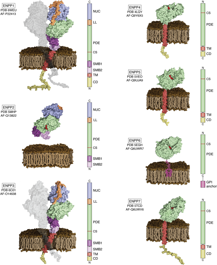
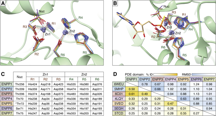
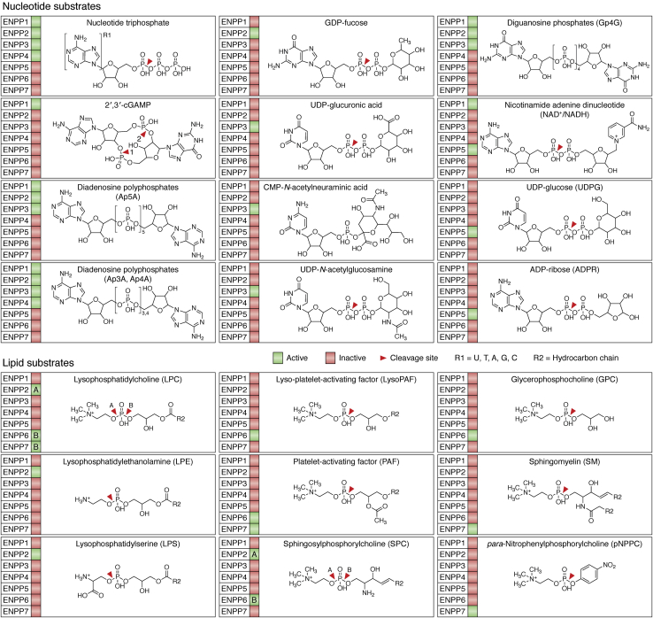
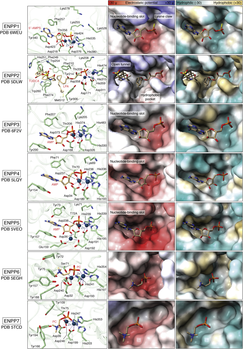
Ecto-nucleotide pyrophosphatase/phosphodiesterase (ENPP) family members (ENPP1-7) have been implicated in key biological and pathophysiological processes, including nucleotide and phospholipid signaling, bone mineralization, fibrotic diseases, and tumor-associated immune cell infiltration. ENPPs are single-pass transmembrane ecto-enzymes, with notable exceptions of ENPP2 (Autotaxin) and ENNP6, which are secreted and glycosylphosphatidylinositol (GPI)-anchored, respectively. ENNP1 and ENNP2 are the best characterized and functionally the most interesting members. Here, we review the structural features of ENPP1-7 to understand how they evolved to accommodate specific substrates and mediate different biological activities. ENPPs are defined by a conserved phosphodiesterase (PDE) domain. In ENPP1-3, the PDE domain is flanked by two N-terminal somatomedin B-like domains and a C-terminal inactive nuclease domain that confers structural stability, whereas ENPP4-7 only possess the PDE domain. Structural differences in the substrate-binding site endow each protein with unique characteristics. Thus, ENPP1, ENPP3, ENPP4, and ENPP5 hydrolyze nucleotides, whereas ENPP2, ENPP6, and ENNP7 evolved as phospholipases through adaptions in the catalytic domain. These adaptations explain the different biological and pathophysiological functions of individual members. Understanding the ENPP members as a whole advances our insights into common mechanisms, highlights their functional diversity, and helps to explore new biological roles.

Keywords: autotaxin; cancer; drug development; mineralization; phosphodiesterases; phospholipase; pyrophosphate; signaling; structure–function.


Conflict of interest statement

Conflict of interest The authors declare that they have no conflicts of interest with the contents of this article.

Figures

Figure 1
Figure 1
Overview of ENPP1–7 structures and domain architecture. The structures are presented as surface models based on experimental findings and AlphaFold models (87). The ENPP1–3 multidomain subgroup is to the left, and the single-domain ENPP4–7 subgroup to the right. All family members are attached to the cell surface through a transmembrane domain, except for secreted ENPP2 and GPI-anchored ENPP6. Next to each structure, the colored bar represents the location of the different domains in the corresponding sequences. All pictures were generated in UCSF ChimeraX 1.2.5 (88), and the scenes were created and rendered using Blender 2.93.5 (https://www.blender.org/). PDE, Phosphodiesterase domain; CS, Catalytic site; SMB1/2, somatomedin like domain 1/2; NUC, Nuclease-like domain; LL, lasso loop; TM, transmembrane domain; CD, cytoplasmic domain; PDB files used: ENPP1: 6WEU, AF-P22413; ENPP2: 5MHP, AF-Q13822; ENPP3: 6C01, AF-O14638; ENPP4: 4LQY, AF-Q9Y6X5; ENPP5: 5VEO, AF-Q9UJA9; ENPP6: 5EGH, AF-Q6UWR7; and ENPP7: 5TCD, AF-Q6UWV6.
Figure 2
Figure 2
The conserved catalytic site of ENPP1–7.A, the zinc ions and the seven residues implicated in catalysis are shown for each ENPP with a different color; all structures have been superimposed in ChimeraX 1.2.5 (88), and the green ribbon model represents ENPP1; Nuc, nucleophile, R1–3, residues coordinating Zn1, R4–6, residues coordinating Zn2 (B) a different orientation of A, also showing binding of adenosine-5′-thio-monophosphate to better depict the substrate orientation. C, summary of the seven conserved residues involved in catalysis; ∗this residue was mutated to Alanine in the crystal structure. D, comparison of the catalytic PDE domains of all seven members for the human ENPPs: the sequence identity (yellow; saturation highlights highly conserved sequences) after multiple sequence alignment using ClustalW (25) is shown to the bottom half; the overall root mean square distance deviations (blue; saturation highlights more similar structures) after flexible structural superposition using RAPIDO (26) are shown; the mouse structure was used for ENNP6.
Figure 3
Figure 3
Substrates of ENPP1–7 divided into nucleotide-like and lipid-containing substrates. For each substrate, family members that have been shown to process these substrates in vitro or in vivo are highlighted in green; red suggests either that the substrate cannot be processed or data are not available. The red arrow-head indicates the scissile bond.
Figure 4
Figure 4
Substrate recognition in the catalytic site. The first column shows the best characterized substrate for each family member as a stick model (carbons: creme; oxygens: red; nitrogen: blue; phosphate: orange) and a ribbon model of the corresponding structure (green) as well as the residues involved in substrate recognition and catalysis (green carbons; other atoms as for the substrate) and the zinc ions (blue). The second column shows the surface colored by electrostatic potential for each structure, and the same substrate as in the previous column as a stick model; the last column shows the surface colored by hydrophobicity. All pictures and calculations were generated with UCSF ChimeraX 1.2.5 (88) PDB files: 6WEU, 5DLW, 6F2V, 5LQY, 5VEO, 5EGH and 5TCD.

References

    1. Takahashi T., Old L.J., Boyse E.A. Surface alloantigens of plasma cells. J. Exp. Med. 1970;131:1325–1341. - PMC - PubMed
    1. van Driel I.R., Goding J.W. Plasma cell membrane glycoprotein PC-1. Primary structure deduced from cDNA clones. J. Biol. Chem. 1987;262:4882–4887. - PubMed
    1. Goding J.W., Terkeltaub R., Maurice M., Deterre P., Sali A., Belli S.I. Ecto-phosphodiesterase/pyrophosphatase of lymphocytes and non-lymphoid cells: Structure and function of the PC-1 family. Immunol. Rev. 1998;161:11–26. - PubMed
    1. Bollen M., Gijsbers R., Ceulemans H., Stalmans W., Stefan C. Nucleotide pyrophosphatases/phosphodiesterases on the move. Crit. Rev. Biochem. Mol. Biol. 2000;35:393–432. - PubMed
    1. Tokumura A., Majima E., Kariya Y., Tominaga K., Kogure K., Yasuda K., Fukuzawa K. Identification of human plasma lysophospholipase D, a lysophosphatidic acid-producing enzyme, as autotaxin, a multifunctional phosphodiesterase. J. Biol. Chem. 2002;277:39436–39442. - PubMed

MeSH terms