

Regional brain atrophy and cognitive decline depend on definition of subjective cognitive decline
- PMID: 34959049
- PMCID: PMC8718726
- DOI: 10.1016/j.nicl.2021.102923
Regional brain atrophy and cognitive decline depend on definition of subjective cognitive decline
Abstract
Background: People with subjective cognitive decline (SCD) may be at increased risk for Alzheimer's disease (AD). However, not all studies have observed this increased risk. This project examined whether four common methods of defining SCD yields different patterns of atrophy and future cognitive decline between cognitively normal older adults with (SCD+ ) and without SCD (SCD-).
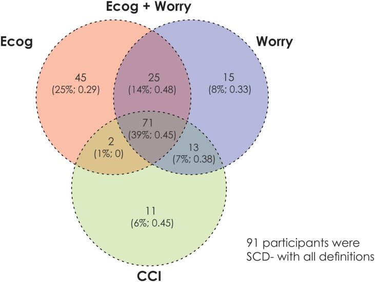
Methods: Data from 273 Alzheimer's Disease Neuroimaging Initiative cognitively normal older adults were examined. To operationalize SCD we used four common methods: Cognitive Change Index (CCI), Everyday Cognition Scale (ECog), ECog + Worry, and Worry. Voxel-based logistic regressions were applied to deformation-based morphology results to determine if regional atrophy between SCD- and SCD+ differed by SCD definition. Linear mixed-effects models were used to evaluate differences in future cognitive decline.
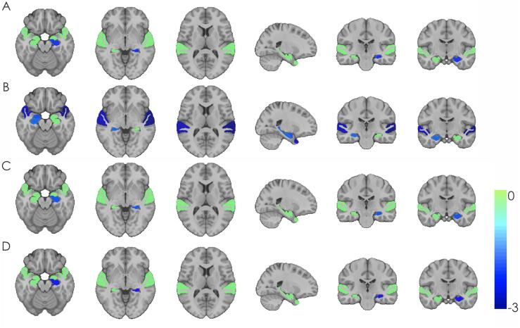
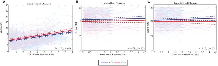
Results: Results varied between the four methods of defining SCD. Left hippocampal grading was more similar to AD in SCD+ than SCD- when using the CCI (p = .041) and Worry (p = .021) definitions. The right (p=.008) and left (p=.003) superior temporal regions had smaller volumes in SCD+ than SCD-, but only with the ECog. SCD+ was associated with greater future cognitive decline measured by Alzheimer's Disease Assessment Scale, but only with the CCI definition. In contrast, only the ECog definition of SCD was associated with future decline on the Montreal Cognitive Assessment.
Conclusion: These findings suggest that the various methods used to differentiate between SCD- and SCD+ influence whether volume differences and findings of cognitive decline are observed between groups in this retrospective analysis.
Keywords: Alzheimer’s disease; Cognitive decline; Deformation based morphometry; Magnetic resonance imaging; Regional atrophy; Subjective cognitive decline.
Copyright © 2021 The Authors. Published by Elsevier Inc. All rights reserved.
Figures
References
-
- Amariglio R.E., Donohue M.C., Marshall G.A., Rentz D.M., Salmon D.P., Ferris S.H., Karantzoulis S., Aisen P.S., Sperling R.A. Tracking early decline in cognitive function in older individuals at risk for Alzheimer disease dementia: the Alzheimer’s Disease Cooperative Study Cognitive Function Instrument. JAMA Neurol. 2015;72(4):446. doi: 10.1001/jamaneurol.2014.3375. - DOI - PMC - PubMed
-
- Alzheimer’s Association, 2021. What is Alzheimer’s disease. Retrieved from https://www.alz.org/alzheimers-dementia/what-is-alzheimers.
-
- Cantero J.L., Iglesias J.E., Van Leemput K., Atienza M. Regional hippocampal atrophy and higher levels of plasma amyloid-beta are associated with subjective memory complaints in nondemented elderly subjects. J. Gerontol. Ser. A: Biomed. Sci. Med. Sci. 2016;71(9):1210–1215. doi: 10.1093/gerona/glw022. - DOI - PMC - PubMed
-
- Chou Y.Y., Leporé N., Avedissian C., Madsen S.K., Parikshak N., Hua X., Shaw L.M., Trojanowski J.Q., Weiner M.W., Toga A.W., Thompson P.M. Mapping correlations between ventricular expansion and CSF amyloid and tau biomarkers in 240 subjects with Alzheimer’s disease, mild cognitive impairment and elderly controls. Neuroimage. 2009;46(2):394–410. doi: 10.1016/j.neuroimage.2009.02.015. - DOI - PMC - PubMed
Publication types
MeSH terms
Grants and funding
LinkOut - more resources
Full Text Sources
Medical
