

Evaluation of Ruthenium-Based Assemblies as Carriers of Photosensitizers to Treat Rheumatoid Arthritis by Photodynamic Therapy
- PMID: 34959385
- PMCID: PMC8706357
- DOI: 10.3390/pharmaceutics13122104
Evaluation of Ruthenium-Based Assemblies as Carriers of Photosensitizers to Treat Rheumatoid Arthritis by Photodynamic Therapy
Abstract
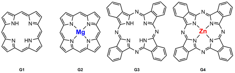
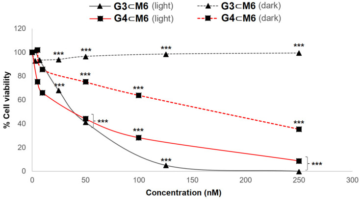
For the first time, ruthenium-based assemblies have been used as carriers for photosensitizers in the treatment of rheumatoid arthritis by photodynamic therapy (PDT). These metallacages are totally soluble in physiological media and can transport photosensitizers (PS) in their cavity. After an incubation period, the PS is released in the cytoplasm and irradiation can take place. This strategy allows photosensitizers with low or null solubility in biological media to be evaluated as PDT agents in rheumatoid arthritis. The systems in which 21H,23H-porphine and 29H,31H-phthalocyanine are encapsulated show excellent photocytotoxicity and no toxicity in the dark. On the other hand, systems in which metalated derivatives such as Mg(II)-porphine and Zn(II)-phthalocyanine are used show good photocytotoxicity, but to a lesser extent than the previous two. Furthermore, the presence of Zn(II)-phthalocyanine significantly increases the toxicity of the system. Overall, fifteen different host-guest systems have been evaluated, and based on the results obtained, they show high potential for treating rheumatoid arthritis by PDT.
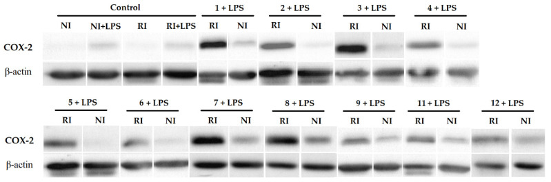
Keywords: COX-2; arene ruthenium complexes; drug delivery; host–guest system; photodynamic therapy; photosensitizer; rheumatoid arthritis.
Conflict of interest statement
The authors declare no conflict of interest.
Figures


References
-
- Roux C.H., Saraux A., Le Bihan E., Fardellone P., Guggenbuhl P., Fautrel B., Masson C., Chary-Valckenaere I., Cantagrel A., Juvin R., et al. Rheumatoid arthritis and spondyloarthropathies: Geographical variations in prevalence in France. J. Rheumatol. 2007;34:117–122. - PubMed
-
- Kerkman P.F., Fabre E., van der Voort E.I., Zaldumbide A., Rombouts Y., Rispens T., Wolbink G., Hoeben R.C., Spits H., Baeten D.L., et al. Identification and characterization of citrullinated antigen-specific B cells in peripheral blood of patients with rheumatoid arthritis. Ann. Rheum. Dis. 2016;75:1170–1176. doi: 10.1136/annrheumdis-2014-207182. - DOI - PubMed
Grants and funding
LinkOut - more resources
Full Text Sources
Research Materials
