Review
. 2021 Dec 5;10(12):1583.
doi: 10.3390/pathogens10121583.

Host Range, Biology, and Species Specificity of Seven-Segmented Influenza Viruses-A Comparative Review on Influenza C and D

Affiliations
Review

Host Range, Biology, and Species Specificity of Seven-Segmented Influenza Viruses-A Comparative Review on Influenza C and D

Chithra C Sreenivasan et al. Pathogens. .

Abstract

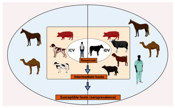
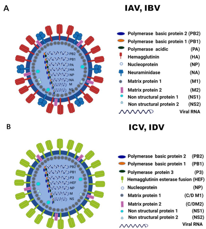
Other than genome structure, influenza C (ICV), and D (IDV) viruses with seven-segmented genomes are biologically different from the eight-segmented influenza A (IAV), and B (IBV) viruses concerning the presence of hemagglutinin-esterase fusion protein, which combines the function of hemagglutinin and neuraminidase responsible for receptor-binding, fusion, and receptor-destroying enzymatic activities, respectively. Whereas ICV with humans as primary hosts emerged nearly 74 years ago, IDV, a distant relative of ICV, was isolated in 2011, with bovines as the primary host. Despite its initial emergence in swine, IDV has turned out to be a transboundary bovine pathogen and a broader host range, similar to influenza A viruses (IAV). The receptor specificities of ICV and IDV determine the host range and the species specificity. The recent findings of the presence of the IDV genome in the human respiratory sample, and high traffic human environments indicate its public health significance. Conversely, the presence of ICV in pigs and cattle also raises the possibility of gene segment interactions/virus reassortment between ICV and IDV where these viruses co-exist. This review is a holistic approach to discuss the ecology of seven-segmented influenza viruses by focusing on what is known so far on the host range, seroepidemiology, biology, receptor, phylodynamics, species specificity, and cross-species transmission of the ICV and IDV.

Keywords: biology; eight-segmented; host range; influenza C; influenza D; receptor 9; seven-segmented; species specificity; structure; transmission.


Conflict of interest statement

The authors declare no conflict of interest.

Figures

Figure 1
Figure 1
Timeline of natural infections of the influenza C and D viruses based on the chronological order of occurrence. The key events of ICV (orange flags) and IDV (green flags) were described on the top and bottom of the year bar, respectively. The right-facing flags (dark orange and dark green) represent the events of virus isolation and the left-facing flags with dotted lines (light orange and light green) denote the events of seroprevalence. Following the first detection of IDV in 2011–2012, serological studies using archived bovine and human sera conducted in the USA and Italy found evidence of IDV circulation in 2003–2005, which is also shown in the figure. The year of the event is also marked next to the flag and the species involved is also illustrated. The graphical illustration was created with BioRender.com, accessed 30 November 2021.
Figure 2
Figure 2
The host ecology of ICV and IDV. The principal hosts/reservoir animals of ICV (humans) and IDV (bovines) have been described in the innermost (white) oval space, followed by the intermediate hosts in the (light orange) middle space based on the natural infection, ICV: swine, canine, bovines; IDV: swine, bovines, caprine, bubaline, etc. The susceptible hosts are shown based on serological information in the outermost (light blue) space. Equines and camelids are susceptible hosts for both ICV and IDV, whereas humans are susceptible to IDV. The graphic image was created with BioRender.com, accessed 30 October 2021.
Figure 3
Figure 3
A synopsis of the tissue tropism demonstrated by the seven-segmented viruses, ICV, and IDV in different experimental animals based on the literature review. The illustration was created using Biorender.com, accessed 30 October 2021.
Figure 4
Figure 4
Phylogenetic tree of the complete protein-coding nucleotide sequences of the ICV-HEF. The evolutionary history of ICV-HEF was inferred using Maximum-likelihood analysis in MEGA X [136]. Tamura 3 parameter model + Gamma Distributed (G) [137] model was used with a bootstrapping of 1000 replicates for the analysis. The analyses involved 94 sequences of ICV strains of human, bovine, and swine origin. Different lineages of ICV are color-coded and marked as follows. Green open diamond—C/Sao Paulo; purple open inverted triangle—C/Aichi; Cyan filled circle—C/Kanagawa; Orange open square—C/Yamagata; Blue open circle—C/Taylor; Red open triangle—C/Mississippi. The strains of bovine ICV (Red filled triangle) and swine ICVs (Orange filled square) belonged to C/Mississippi and C/Yamagata, respectively. A scale representing evolutionary distance that indicates the number of nucleotide substitutions per site is also shown. Bootstrap values are shown above branches to the left of major nodes.
Figure 5
Figure 5
Phylogenetic tree of the complete protein-coding nucleotide sequences of the IDV-HEF. Phylogeny was inferred using maximum-likelihood analysis in MEGA X [136], with a bootstrapping of 1000 replicates. The tree was derived using Hasegawa-Kishino-Yano model + Gamma Distributed (G) [HKY + G] model. The analyses involved 38 sequences of IDV strains of bovine and swine origin from different geographic locations. Different lineages of IDV are color-coded and marked as follows: D/OK (Light Orange filled triangle), D/660 (green open square), D/CA2019 (Red filled square), D/Yama2019 (brown open circle), and D/Yama2016 (Purple filled circle). A scale representing evolutionary distance that indicates the number of nucleotide substitutions per site is also shown. Bootstrap values are shown above branches to the left of major nodes.
Figure 6
Figure 6
The structural differences of eight segmented influenza viruses and seven segmented influenza viruses. The different proteins are marked with a description on the right. (A) Eight-segmented influenza viruses (IAV, and IBV) with separately encoded Hemagglutinin (HA) and neuraminidase (NA) proteins for receptor binding and destroying functions, respectively. (B) Seven-segmented influenza viruses with Hemagglutinin–esterase fusion protein that combines the dual function of HA and NA. The graphical illustration was created using Biorender.com, accessed 30 October 2021.
Figure 7
Figure 7
Broad differences of N-glycosylation sites of the hemagglutinin esterase fusion protein between influenza C (A) and D viruses (B). The reference genomes used for the structure modeling are C/Johannesburg/1/66 (A) and D/swine/Oklahoma/1334/2011 (B). High confident N-glycosylation sites (Asn having >40 angstrom2 side-chain accessible surface area) were predicted by GLYCAM website. MAN-3 was added to each N-glycosylation site (colored green). HA1 and HA2 were colored light blue and wheat, respectively.

References

    1. Potter C.W. A history of influenza. J. Appl. Microbiol. 2001;91:572–579. doi: 10.1046/j.1365-2672.2001.01492.x. - DOI - PubMed
    1. Fields Virology: Emerging Viruses. Volume 1 Wolters Kluwer; Philadelphia, PA, USA: 2021.
    1. Callan R.J., Early G., Kida H., Hinshaw V.S. The appearance of H3 influenza viruses in seals. J. Gen. Virol. 1995;76:199–203. doi: 10.1099/0022-1317-76-1-199. - DOI - PubMed
    1. Kawano J., Onta T., Kida H., Yanagawa R. Distribution of antibodies in animals against influenza B and C viruses. Jpn. J. Vet. Res. 1978;26:74–80. - PubMed
    1. Chang C.P., New A.E., Taylor J.F., Chiang H.S. Influenza virus isolations from dogs during a human epidemic in Taiwan. Int. J. Zoonoses. 1976;3:61–64. - PubMed

LinkOut - more resources