Review
. 2021 Nov 23;14(12):1210.
doi: 10.3390/ph14121210.

Antiviral Activities of Eucalyptus Essential Oils: Their Effectiveness as Therapeutic Targets against Human Viruses

Affiliations
Review

Antiviral Activities of Eucalyptus Essential Oils: Their Effectiveness as Therapeutic Targets against Human Viruses

Daniel Mieres-Castro et al. Pharmaceuticals (Basel). .

Abstract

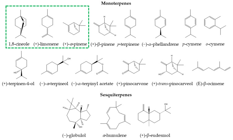
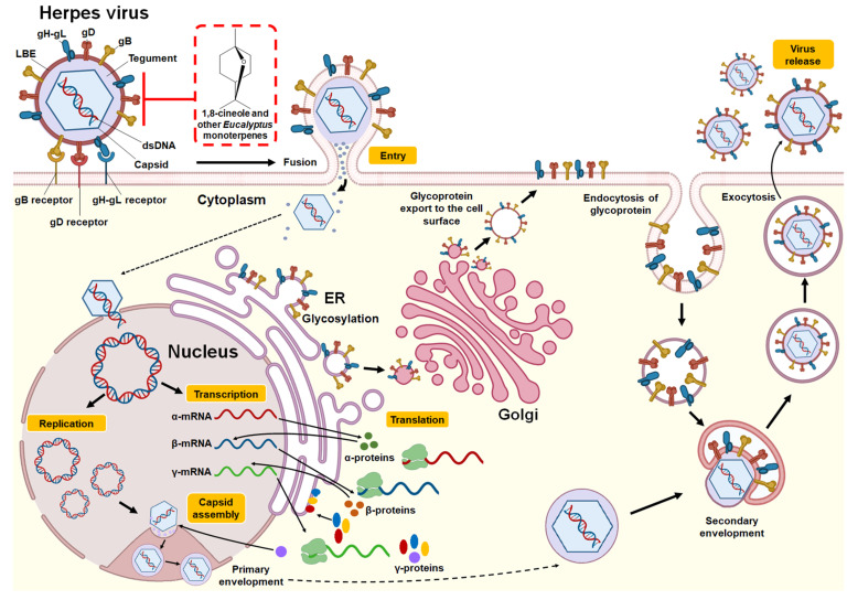
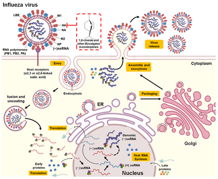
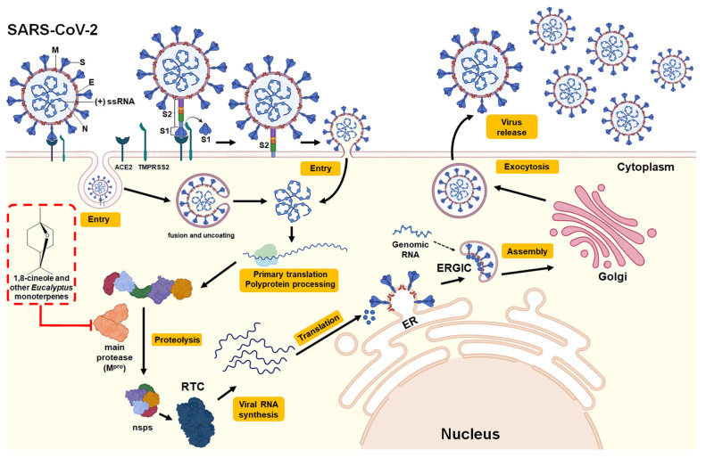
Given the limited therapeutic management of infectious diseases caused by viruses, such as influenza and SARS-CoV-2, the medicinal use of essential oils obtained from Eucalyptus trees has emerged as an antiviral alternative, either as a complement to the treatment of symptoms caused by infection or to exert effects on possible pharmacological targets of viruses. This review gathers and discusses the main findings on the emerging role and effectiveness of Eucalyptus essential oil as an antiviral agent. Studies have shown that Eucalyptus essential oil and its major monoterpenes have enormous potential for preventing and treating infectious diseases caused by viruses. The main molecular mechanisms involved in the antiviral activity are direct inactivation, that is, by the direct binding of monoterpenes with free viruses, particularly with viral proteins involved in the entry and penetration of the host cell, thus avoiding viral infection. Furthermore, this review addresses the coadministration of essential oil and available vaccines to increase protection against different viruses, in addition to the use of essential oil as a complementary treatment of symptoms caused by viruses, where Eucalyptus essential oil exerts anti-inflammatory, mucolytic, and spasmolytic effects in the attenuation of inflammatory responses caused by viruses, in particular respiratory diseases.

Keywords: 1,8-cineole; Eucalyptus essential oil; H1N1 influenza virus; SARS-CoV-2; antiviral therapy; herpes simplex virus.


Conflict of interest statement

The authors declare no conflict of interest.

Figures

Figure 1
Figure 1
Chemical structure of main monoterpenes and sesquiterpenes present in Eucalyptus essential oils for medicinal use. The green box indicates constituents present in natural medicines with Myrtol® standardized.
Figure 2
Figure 2
Structure and replication cycle of Herpes virus. According to Karasneh et al. [46] and Lussignol et al. [47], the Herpes virus consists of 7 structural glycoproteins (gB, gC, gD, gH, gK, gL and gM) present in the lipid bilayer envelope (LBE). However, only four of these glycoproteins (gB, gD, gH, and gL) are necessary and sufficient to allow the fusion of the virus with the plasma membrane of the host cell (shown in the illustration). It has a relatively large, double-stranded, linear DNA genome surrounded by an icosahedral capsid. This, in turn, is surrounded by an integument that contains between 15 and 20 proteins and is in direct contact with the LBE. The herpes virus replication cycle begins when the gB, gD, and gH-gL glycoproteins bind to their receptors in the host cell (gB receptors: PILRα (HSV-1), MAG, NMHC-IIA; gD receptors: HVEM, Nectin-1/Nectin-2, 3-OS HS (HSV-1); gH-gL receptors: αvβ3 integrin). This allows the LBE of the virus to fuse with the plasma membrane or endocytosis, releasing the capsid and integument into the cytoplasm. Using the microtubule network, the nucleocapsid is transported to the nuclear pore, where the viral genome is released into the nucleus and circularized. Viral DNA serves as a template for RNA polymerase II, which leads to the production of mRNA, expressed in three successive and coordinated phases. The mRNAs are translated in the cytoplasm into different viral proteins, including immediate-early (α-proteins), early (β-proteins), and late (γ-proteins) proteins. Most of the late gene products contribute to the formation of the viral particle. Packaging of DNA into preassembled capsids takes place in the nucleus. This is followed by a primary envelope of the capsid by budding through the inner nuclear membrane. The envelope of the perinuclear virions then fuses with the outer nuclear membrane to release naked capsids into the cytoplasm (de-envelopment). The envelope proteins are glycosylated in the endoplasmic reticulum (ER) and then move by transport vesicle from the ER to the Golgi apparatus and finally to the cell plasma membrane. Tegumented capsids acquire a “second” final envelope to become virions from post-Golgi membrane compartments. A role for autophagic membranes in virion envelope and release has been proposed for some herpes viruses. Once formed, virions are transported to the cell surface within small vesicles using exocytosis machinery and released from cells. The red box indicates the potential mechanism of action against the Herpes virus by 1,8-cineole and other monoterpenes present in Eucalyptus essential oils by binding and inhibiting the glycoproteins gB, gD, and gH-gL and thus inhibiting the binding of the virus with its receptors and subsequent fusion of LBE with the host cell.
Figure 3
Figure 3
Structure and replication cycle of the Influenza A virus. According to Neumann et al. [57] and Shi et al. [58], Influenza A virus consists of structural proteins present in the lipid bilayer envelope (LBE), including hemagglutinin (HA), neuraminidase (NA), the matrix protein M1, and ion channel protein M2. It has a negative-sense single-stranded RNA (−ssRNA) genome that contains eight gene segments that encode 16 proteins (although not all influenza viruses express all 16 proteins). Its genomic RNA is encapsulated by nucleoprotein (NP) and components of the RNA-dependent RNA polymerase complex (PB1, PB2, and PA). The replication cycle of the influenza A virus is initiated by the binding of the virus HA to the sialylated receptors (α2,3 or α2,6-linked sialic acid) on the surface of the host cell. This allows endocytosis-mediated entry of the virus. Following the fusion of the virus and host cell membranes, uncoating occurs and the release of viral RNA into the cytoplasm. Subsequently, the −ssRNA genome (noncoding) is transported to the nucleus, where replication and transcription into coding RNA (+ssRNA) occur. Messenger RNAs are exported to the cytoplasm for translation. The early viral proteins, that is, those necessary for replication and transcription (NP, NS1, PA, PB1, PB2, PB1-F2), are transported back to the nucleus. After synthesis in the cytoplasm, NP proteins stabilize the −ssRNA in the nucleus. Genomic RNA with RNA polymerase, NP, matrix proteins, and packaging proteins are exported from the nucleus to the cytoplasm with the help of M1 and NS2 proteins (late viral proteins). The envelope proteins produced in the endoplasmic reticulum (ER) move through the transport vesicle from the ER to the Golgi apparatus and then to the plasma membrane. Finally, the genomic RNA and the viral protein complex are packaged into progeny viruses as they emerge from the cell membrane by exocytosis. The red box indicates the potential mechanism of action against influenza A virus by 1,8-cineole and other monoterpenes present in Eucalyptus essential oil by binding and inhibiting the hemagglutinin protein and thus inhibiting the binding of the virus with its receptor and subsequent entry into the host cell.
Figure 4
Figure 4
Structure and viral replication cycle of SARS-CoV-2. According to V’kovski et al. [64], SARS-CoV-2 consists of structural proteins including Spike (S), Membrane (M), Nucleocapsid (N), and, for some beta coronaviruses, hemagglutinin esterase (not shown). The positive-sense single-stranded RNA (+ssRNA) genome is encapsulated by N, while M and E are incorporated into the viral particle during the assembly process. The replication cycle begins with the arrival of the SARS-CoV-2 virus to the target cell. The S viral protein binds to its receptor in the cell, the angiotensin-converting enzyme 2 (ACE2). After receptor binding, the S protein is cleaved by the cell surface serine protease TMPRSS2, forming two subunits, the S1 subunit containing the receptor-binding domain (RBD) and the S2 subunit containing the binding peptide to the fusion protein present in the cell membrane, allowing the entry of the virus into the host cell, either through the formation of an endosome or by the fusion of the viral envelope. Following the fusion of the virus and host cell membranes, the uncoating occurs and the release of viral RNA into the cytoplasm to initiate the translation of coterminal polyproteins (pp1a/ab), which carry out the replication of the viral genome. After translation of viral RNA into polyproteins, the major protease (Mpro), a homodimeric cysteine protease, self-cleaves in order to cleave polyproteins into nonstructural proteins (nsps). Several nsp proteins interact with nsp12 (also called RNA-dependent RNA polymerase (RdRp)) to form the replicase–transcriptase complex (RTC), which is responsible for the synthesis of the full-length viral genome (replication) and subgenomic RNA (transcription). Viral structural proteins are expressed and transferred to the endoplasmic reticulum (ER). Genomic RNA encapsulated in protein N is translocated with structural proteins in the ER-Golgi intermediate compartment (ERGIC) to form new viral particles. Finally, the new virions are secreted from the infected cell by exocytosis. The red box indicates the potential mechanism of action against SARS-CoV-2 by 1,8-cineole and other monoterpenes present in Eucalyptus essential oils by inhibiting Mpro (binding to the active site), thus inhibiting proteolysis of viral polyproteins necessary for virus replication.

References

    1. Chandorkar N., Tambe S., Amin P., Madankar C. A systematic and comprehensive review on current understanding of the pharmacological actions, molecular mechanisms, and clinical implications of the genus Eucalyptus. Phyto. Plus. 2021;1:100089. doi: 10.1016/j.phyplu.2021.100089. - DOI
    1. Valenzuela C.E., Ballesta P., Ahmar S., Fiaz S., Heidari P., Maldonado C., Mora-Poblete F. Haplotype- and SNP-Based GWAS for Growth and Wood Quality Traits in Eucalyptus cladocalyx Trees under Arid Conditions. Plants. 2021;10:148. doi: 10.3390/plants10010148. - DOI - PMC - PubMed
    1. Ballesta P., Serra N., Guerra F.P., Hasbún R., Mora F. Genomic Prediction of Growth and Stem Quality Traits in Eucalyptus globulus Labill. at Its Southernmost Distribution Limit in Chile. Forests. 2018;9:779. doi: 10.3390/f9120779. - DOI
    1. Mora F., Ballesta P., Serra N. Bayesian analysis of growth, stem straightness and branching quality in full-sib families of Eucalyptus globulus. Bragantia. 2019;78:328–336. doi: 10.1590/1678-4499.20180317. - DOI
    1. Ballesta P., Bush D., Silva F.F., Mora F. Genomic Predictions Using Low-Density SNP Markers, Pedigree and GWAS Information: A Case Study with the Non-Model Species Eucalyptus cladocalyx. Plants. 2020;9:99. doi: 10.3390/plants9010099. - DOI - PMC - PubMed

LinkOut - more resources