. 2021 Dec 16;14(12):1313.
doi: 10.3390/ph14121313.

Promising Antiviral Activity of Agrimonia pilosa Phytochemicals against Severe Acute Respiratory Syndrome Coronavirus 2 Supported with In Vivo Mice Study

Affiliations

Promising Antiviral Activity of Agrimonia pilosa Phytochemicals against Severe Acute Respiratory Syndrome Coronavirus 2 Supported with In Vivo Mice Study

Nashwah G M Attallah et al. Pharmaceuticals (Basel). .

Abstract

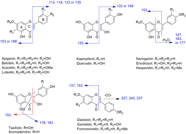
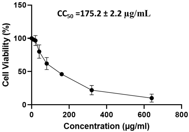
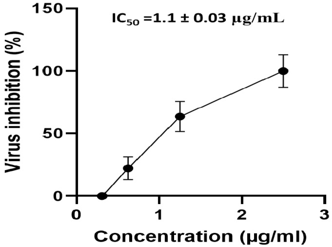
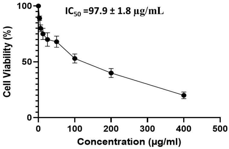
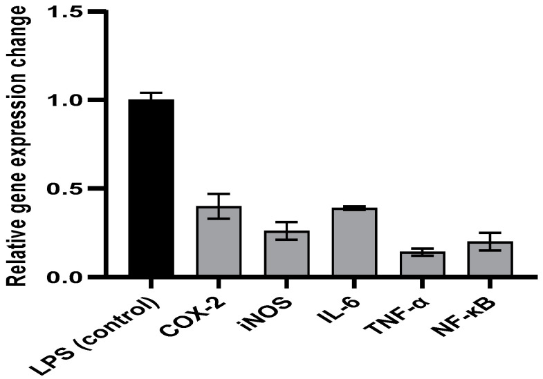
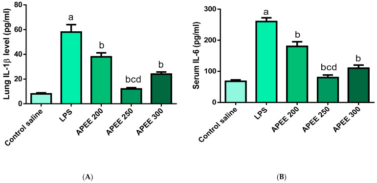
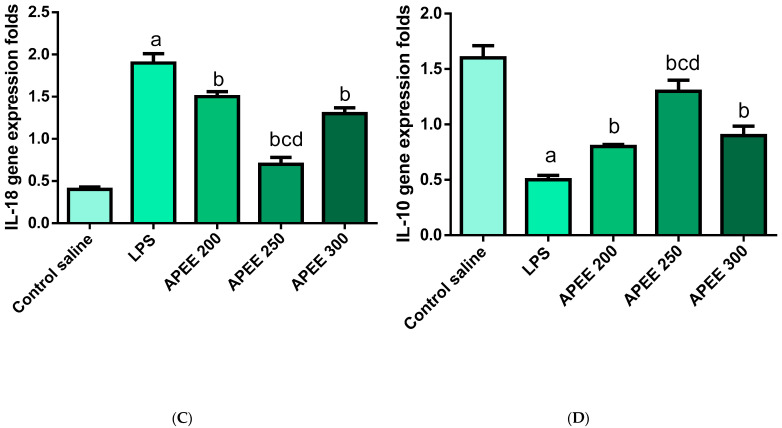
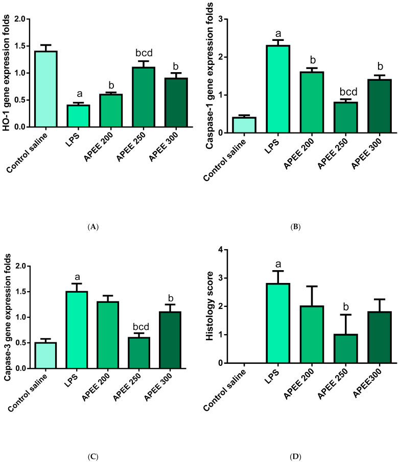
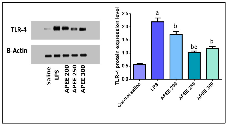
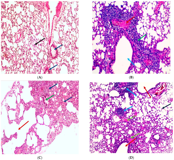
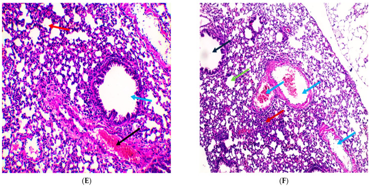
The global emergence of the COVID-19 pandemic caused by severe acute respiratory syndrome coronavirus 2 (SARS-CoV-2) has focused the entire world's attention toward searching for a potential remedy for this disease. Thus, we investigated the antiviral activity of Agrimonia pilosa ethanol extract (APEE) against SARS-CoV-2 and it exhibited a potent antiviral activity with IC50 of 1.1 ± 0.03 µg/mL. Its mechanism of action was elucidated, and it exhibited a virucidal activity and an inhibition of viral adsorption. Moreover, it presented an immunomodulatory activity as it decreased the upregulation of gene expression of COX-2, iNOS, IL-6, TNF-α, and NF-κB in lipopolysaccharide (LPS)-induced peripheral blood mononuclear cells. A comprehensive analysis of the phytochemical fingerprint of APEE was conducted using LC-ESI-MS/MS technique for the first time. We detected 81 compounds and most of them belong to the flavonoid and coumarin classes. Interestingly, isoflavonoids, procyanidins, and anthocyanins were detected for the first time in A. pilosa. Moreover, the antioxidant activity was evidenced in DPPH (IC50 62.80 µg/mL) and ABTS (201.49 mg Trolox equivalents (TE)/mg) radical scavenging, FRAP (60.84 mg TE/mg), and ORAC (306.54 mg TE/g) assays. Furthermore, the protective effect of APEE was investigated in Lipopolysaccharides (LPS)-induced acute lung injury (ALI) in mice. Lung W/D ratio, serum IL-6, IL-18, IL-1β, HO-1, Caspase-1, caspase-3, TLR-4 expression, TAC, NO, MPO activity, and histopathological examination of lung tissues were assessed. APEE induced a marked downregulation in all inflammation, oxidative stress, apoptosis markers, and TLR-4 expression. In addition, it alleviated all histopathological abnormalities confirming the beneficial effects of APEE in ALI. Therefore, APEE could be a potential source for therapeutic compounds that could be investigated, in future preclinical and clinical trials, in the treatment of patients with COVID-19.

Keywords: ALI; LC-ESI-MS/MS; LPS; SARS-CoV-2; TLR-4; immunomodulatory; plaque reduction.


Conflict of interest statement

The authors declare no conflict of interest.

Figures

Figure 1
Figure 1
The structures and fragmentation pattern in positive ion mode for the identified aglycones.
Figure 2
Figure 2
A graph showing the cytotoxicity of APEE on Vero-E6 cells using MTT assay to determine CC50. The results are expressed as mean ± SD as the experiments were performed in three independent triplicates.
Figure 3
Figure 3
A curve showing the effect of APEE different concentrations on the viability of NRC-03-nhCoV. The results are expressed as mean ± SD as the experiments were performed in three independent triplicates.
Figure 4
Figure 4
A graph showing cytotoxicity APEE on PBMCs using MTT to determine IC50. The results are expressed as mean ± SD as the experiments were performed in three independent triplicates.
Figure 5
Figure 5
A chart representing the impact of APEE on the expression of the genes encoding COX-2, iNOS, IL-6, TNF-α, and NF-κB in the LPS-induced PBMCs. The results are expressed as mean ± SD as the experiments were performed in three independent triplicates.
Figure 6
Figure 6
Impact of APEE pre-treatment on (A) Lung IL-1β level, (B) Serum IL-6 level (C) IL-18 gene expression level, (D) IL-10 gene expression level. Acute lung injury was urged by I.P. injection of LPS (10 mg/kg). APEE 200, 250, and 300 were given I.P. 30 min before LPS injection. Results were expressed as mean ± SD (n = 10/group) as the experiments were performed in three independent triplicates. Significant difference vs. a respective control, b respective LPS group, c respective APEE 200 group, d respective APEE 300 group each at p < 0.05.
Figure 6
Figure 6
Impact of APEE pre-treatment on (A) Lung IL-1β level, (B) Serum IL-6 level (C) IL-18 gene expression level, (D) IL-10 gene expression level. Acute lung injury was urged by I.P. injection of LPS (10 mg/kg). APEE 200, 250, and 300 were given I.P. 30 min before LPS injection. Results were expressed as mean ± SD (n = 10/group) as the experiments were performed in three independent triplicates. Significant difference vs. a respective control, b respective LPS group, c respective APEE 200 group, d respective APEE 300 group each at p < 0.05.
Figure 7
Figure 7
Impact of APEE pre-treatment on (A) HO-1 expression level, (B) Caspase-1 expression level (C) Caspase-3 expression level, (D) Lung Histology score. Acute lung injury was urged by I.P. injection of LPS (10 mg/kg). APEE 200, 250, and 300 were given I.P. 30 min before LPS injection. Results were expressed as mean ± SD (n = 10/group) as the experiments were performed in three independent triplicates. Significant difference vs. a respective control, b respective LPS group, c respective APEE 200 group, d respective APEE 300 group each at p < 0.05.
Figure 8
Figure 8
Effect of APEE pre-treatment on the expression of TLR-4 in the lung tissues. The expression levels were measured by western blotting. Acute lung injury was urged by I.P. injection of LPS (10 mg/kg). APEE 200, 250, and 300 were given I.P. 30 min before LPS injection. Results were expressed as mean ± SD (n = 10/group) as the experiments were performed in three independent triplicates. Significant difference vs. a respective control, b respective LPS group, c respective APEE 200 group each at p < 0.05.
Figure 9
Figure 9
Histopathological examination of H&E-stained sections of lung tissue indicates the influence of APEE treatment on LPS-induced ALI. (A) A section in lung of the normal control group indicated normal-sized alveoli separated by fibrous septa (blue arrows) and normal-sized bronchiole (black arrow) (H&E X 100). (B) Section in lung of LPS group showed dilated bronchiole (blue arrow) surrounded by marked chronic inflammation and pneumonia (green arrow) and congested vessels (red arrow) (H&E X 200). (C) Section in lung of LPS group showed dilated destructed alveolar walls (emphysema) (red arrow) surrounded by destructed bronchioles (green arrow) and alveolar congestion with fibrosis (blue arrows) (H&E X 100). (D) Section in lung of APEE 200 treated group showed dilated bronchioles (red arrows) surrounded by decreased interstitial inflammation to moderate degree (blue arrows), congested vessels (green arrows) and decreased emphysema (black arrow) (H&E X 100). (E) Section in lung of APEE 250 treated group showed marked remission of inflammation with average-sized of a bronchiole (blue arrow) surrounded by normal-sized alveoli (red arrow) with few congested vessels (black arrow) (H&E X 200). (F) Section in lung of APEE 300 treated group showed focal inflammation (red arrow) surrounded by average-sized of a bronchiole (black arrow) surrounded by normal-sized alveoli (green arrow) with many congested vessels (blue arrows) (H&E X 100).
Figure 9
Figure 9
Histopathological examination of H&E-stained sections of lung tissue indicates the influence of APEE treatment on LPS-induced ALI. (A) A section in lung of the normal control group indicated normal-sized alveoli separated by fibrous septa (blue arrows) and normal-sized bronchiole (black arrow) (H&E X 100). (B) Section in lung of LPS group showed dilated bronchiole (blue arrow) surrounded by marked chronic inflammation and pneumonia (green arrow) and congested vessels (red arrow) (H&E X 200). (C) Section in lung of LPS group showed dilated destructed alveolar walls (emphysema) (red arrow) surrounded by destructed bronchioles (green arrow) and alveolar congestion with fibrosis (blue arrows) (H&E X 100). (D) Section in lung of APEE 200 treated group showed dilated bronchioles (red arrows) surrounded by decreased interstitial inflammation to moderate degree (blue arrows), congested vessels (green arrows) and decreased emphysema (black arrow) (H&E X 100). (E) Section in lung of APEE 250 treated group showed marked remission of inflammation with average-sized of a bronchiole (blue arrow) surrounded by normal-sized alveoli (red arrow) with few congested vessels (black arrow) (H&E X 200). (F) Section in lung of APEE 300 treated group showed focal inflammation (red arrow) surrounded by average-sized of a bronchiole (black arrow) surrounded by normal-sized alveoli (green arrow) with many congested vessels (blue arrows) (H&E X 100).

References

    1. Zhou P., Yang X.-L., Wang X.-G., Hu B., Zhang L., Zhang W., Si H.-R., Zhu Y., Li B., Huang C.-L. A pneumonia outbreak associated with a new coronavirus of probable bat origin. Nature. 2020;579:270–273. doi: 10.1038/s41586-020-2012-7. - DOI - PMC - PubMed
    1. Huang C., Wang Y., Li X., Ren L., Zhao J., Hu Y., Zhang L., Fan G., Xu J., Gu X. Clinical features of patients infected with 2019 novel coronavirus in Wuhan, China. Lancet. 2020;395:497–506. doi: 10.1016/S0140-6736(20)30183-5. - DOI - PMC - PubMed
    1. Boban M. Novel coronavirus disease (COVID-19) update on epidemiology, pathogenicity, clinical course and treatments. Int. J. Clin. Pract. 2021;75:e13868. doi: 10.1111/ijcp.13868. - DOI - PMC - PubMed
    1. Aguilar R.B., Hardigan P., Mayi B., Sider D., Piotrkowski J., Mehta J.P., Dev J., Seijo Y., Camargo A.L., Andux L. Current understanding of COVID-19 clinical course and investigational treatments. Front. Med. 2020;7:638. doi: 10.3389/fmed.2020.555301. - DOI - PMC - PubMed
    1. Lin S.-N., Rui J., Chen Q.-P., Zhao B., Yu S.-S., Li Z.-Y., Zhao Z.-Y., Wang Y., Zhu Y.-Z., Xu J.-W. Effectiveness of potential antiviral treatments in COVID-19 transmission control: A modelling study. Infect. Dis. Poverty. 2021;10:1–17. doi: 10.1186/s40249-021-00835-2. - DOI - PMC - PubMed