. 2021 Dec 6;13(12):4374.
doi: 10.3390/nu13124374.

Astaxanthin, a Marine Carotenoid, Maintains the Tolerance and Integrity of Adipose Tissue and Contributes to Its Healthy Functions

Affiliations

Astaxanthin, a Marine Carotenoid, Maintains the Tolerance and Integrity of Adipose Tissue and Contributes to Its Healthy Functions

Allah Nawaz et al. Nutrients. .

Abstract

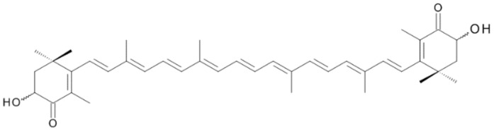
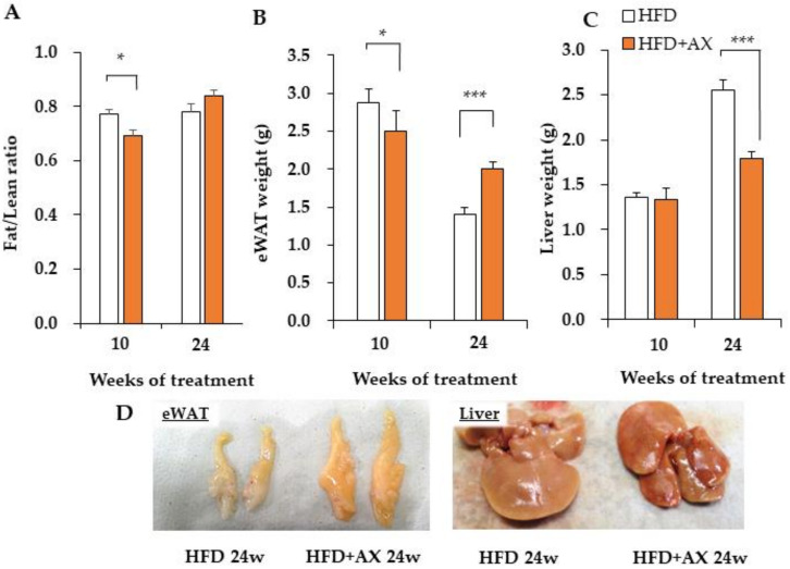
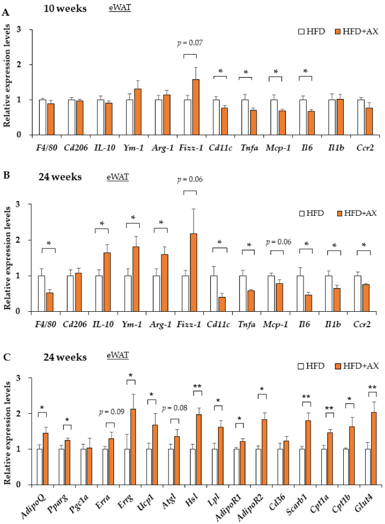
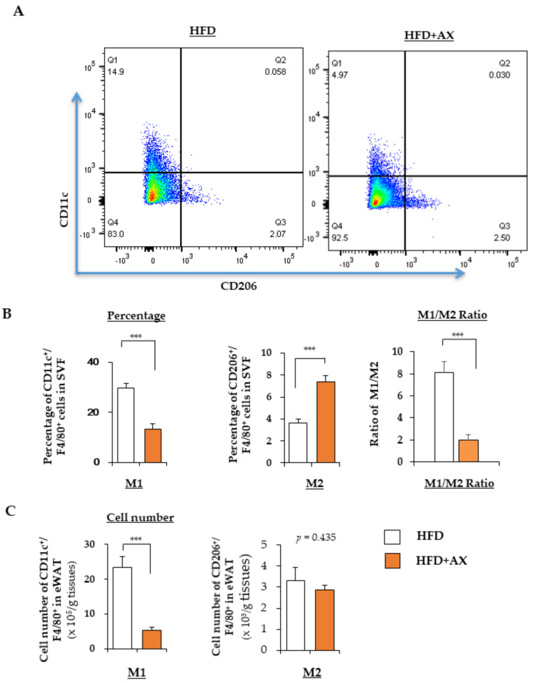
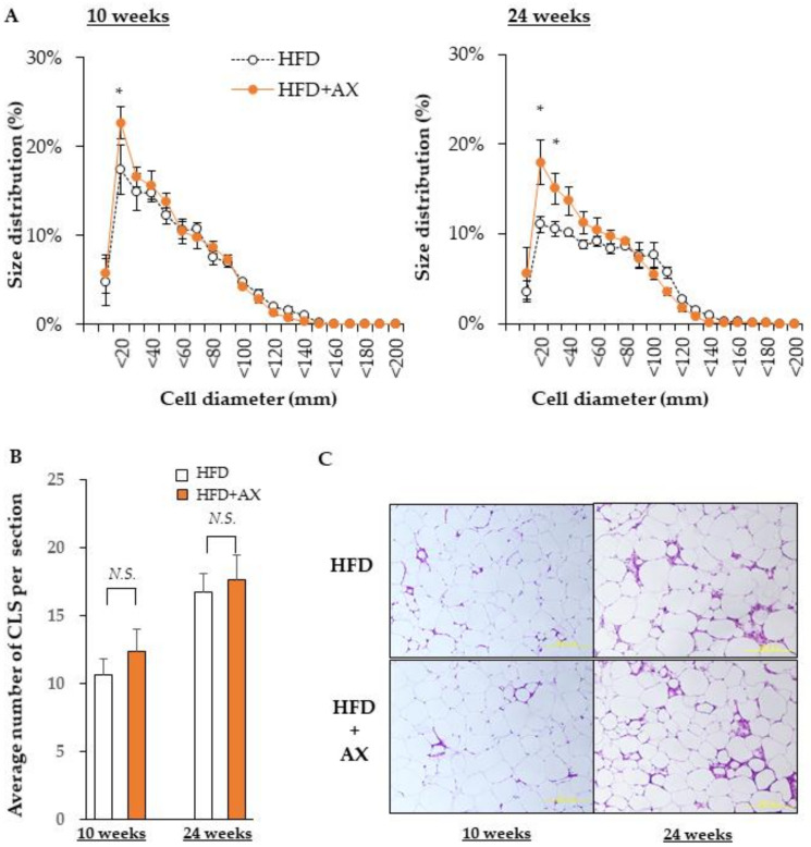
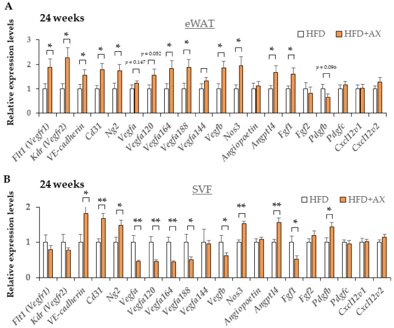
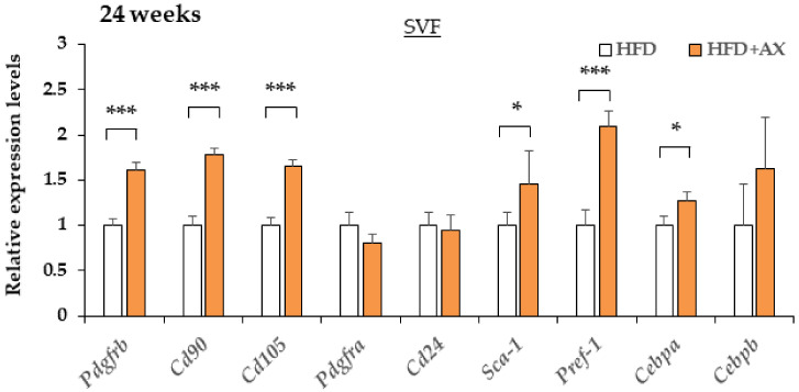
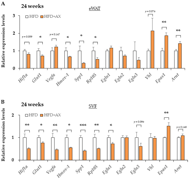
Recently, obesity-induced insulin resistance, type 2 diabetes, and cardiovascular disease have become major social problems. We have previously shown that Astaxanthin (AX), which is a natural antioxidant, significantly ameliorates obesity-induced glucose intolerance and insulin resistance. It is well known that AX is a strong lipophilic antioxidant and has been shown to be beneficial for acute inflammation. However, the actual effects of AX on chronic inflammation in adipose tissue (AT) remain unclear. To observe the effects of AX on AT functions in obese mice, we fed six-week-old male C57BL/6J on high-fat-diet (HFD) supplemented with or without 0.02% of AX for 24 weeks. We determined the effect of AX at 10 and 24 weeks of HFD with or without AX on various parameters including insulin sensitivity, glucose tolerance, inflammation, and mitochondrial function in AT. We found that AX significantly reduced oxidative stress and macrophage infiltration into AT, as well as maintaining healthy AT function. Furthermore, AX prevented pathological AT remodeling probably caused by hypoxia in AT. Collectively, AX treatment exerted anti-inflammatory effects via its antioxidant activity in AT, maintained the vascular structure of AT and preserved the stem cells and progenitor's niche, and enhanced anti-inflammatory hypoxia induction factor-2α-dominant hypoxic response. Through these mechanisms of action, it prevented the pathological remodeling of AT and maintained its integrity.

Keywords: Astaxanthin; adipose tissue macrophages; adipose tissue remodeling; insulin resistance; natural antioxidant; obesity.


Conflict of interest statement

Y.N. is currently employed by Fuji Chemical Industries, Co., Ltd. All other authors declare that there is no duality of interest associated with this manuscript.

Figures

Figure 1
Figure 1
Chemical structure of Astaxanthin (AX).
Figure 2
Figure 2
AX administration changed the body composition and tissue weight of high-fat diet-loaded (HFD) mice. (A) Body composition by NMR, Fat/lean ratio, epididymal adipose tissue (eWAT) weight (B), and liver weight (C) of control mice on a high-fat diet and AX-treated mice at 10 and 24 weeks. (D) Representative images of eWAT and liver tissues. (n = 5–6 per group). All values are represented as means ± S.E.M. * p < 0.05, *** p < 0.001 (HFD vs. HFD + AX). Statistical tests were performed as follows: two-way repeated-measures ANOVA, a post-hoc Tukey-Kramer for each point.
Figure 3
Figure 3
AX administration regulated the gene expression of pro-inflammatory markers and metabolic markers in the eWAT compared to HFD-treated control mice. Gene expression of pro-inflammatory and anti-inflammatory-related marker genes in eWAT of HFD-fed mice either 8 weeks (A) or 24 weeks (B) after AX administration. Gene expression of metabolism markers, including adipokines, lipolysis and energy metabolisms in eWAT of HFD treated for 10 weeks (n = 5–6 per group) (C). All values are presented as the means ± S.E.M. * p < 0.05, ** p < 0.01 (HFD vs. HFD + AX). Statistical analysis was performed using Student’s t-test.
Figure 4
Figure 4
AX administration attenuated the infiltration of M1 macrophages (MΦ) into adipose tissue of HFD-fed mice. (A) Representative flow cytometry analysis of immune cells in eWAT of AX-treated HFD and HFD control mice (n = 6 mice/group). For this, the live cells were gated for CD45+ cells, followed by F4/80+ MΦ, and CD206+ M2 MΦ, and CD11c+ M2 MΦ. The percentages, their M1/M2 MΦ ratio (B) and the cell numbers (C) of M1 (CD11c+) and M2 (CD206+) MΦ relative to F4/80+ cells in the stromal vascular fraction (SVF) of eWAT from mice treated with HFD for 24 weeks. All values are presented as the means ± S.E.M. *** p < 0.001 (HFD vs. HFD + AX). Statistical analysis was performed using Student’s t-test.
Figure 5
Figure 5
AX administration significantly altered the size of adipocytes in eWAT. (A) Diameter of adipocytes calculated from the area of adipocytes. (B) Average numbers of the crown like structures in each Hematoxylin and Eosin (H&E) stained section. (C) Representative H&E stained histological sections (n = 3–4/each group, each sample was measured in at least four sections). All values are represented as means ± S.E.M. * p < 0.05, N.S.: not significant (HFD vs. HFD + AX). Statistical tests were performed as follows: (A) two-way repeated-measures ANOVA, a post-hoc Dunnet’s-test for each point. (B) Student’s t-test.
Figure 6
Figure 6
AX administration partially upregulated the gene expression of vascularization markers in eWAT and their SVF compared to HFD-treated control mice for 24 weeks. Gene expression of vascular marker genes in eWAT (A) and their SVF (B) (n = 5–6/each group). All values are represented as means ± S.E.M. * p < 0.05, ** p < 0.01 (HFD vs. HFD + AX). Statistical tests were performed as follows: Student’s t-test.
Figure 7
Figure 7
AX administration altered the gene expression of adipocyte progenitor cells, and mesenchymal stem cell markers in SVF compared to HFD-treated control mice for 24 weeks. Gene expression of adipose progenitor cells, and stem cell marker genes in SVF from eWAT of mice treated HFD for 24 weeks. (n = 5–6/each group). All values are represented as means ± S.E.M. * p < 0.05, *** p < 0.001 (HFD vs. HFD + AX). Statistical tests were performed as follows: Student’s t-test.
Figure 8
Figure 8
The hypoxic response altered from HIF-1 to HIF-2 dominant in eWAT and its SVF of AX-treated HFD mice compared to HFD control mice for 24 weeks. Gene expression of vascular marker genes in eWAT (A) and their SVF (B). (n = 5–6/each group). All values are represented as means ± S.E.M. * p < 0.05, ** p < 0.01, *** p < 0.001 (HFD vs. HFD+AX). Statistical tests were performed as follows: Student’s t-test.

References

    1. Friedman J.M. Obesity in the new millennium. Nature. 2000;404:632–634. doi: 10.1038/35007504. - DOI - PubMed
    1. Cho N.H., Shaw J.E., Karuranga S., Huang Y., da Rocha Fernandes J.D., Ohlrogge A.W., Malanda B. IDF Diabetes Atlas: Global estimates of diabetes prevalence for 2017 and projections for 2045. Diabetes Res. Clin. Pract. 2018;138:271–281. doi: 10.1016/j.diabres.2018.02.023. - DOI - PubMed
    1. Reinehr T. Type 2 diabetes mellitus in children and adolescents. World J. Diabetes. 2013;4:270–281. doi: 10.4239/wjd.v4.i6.270. - DOI - PMC - PubMed
    1. Chait A., den Hartigh L.J. Adipose Tissue Distribution, Inflammation and Its Metabolic Consequences, Including Diabetes and Cardiovascular Disease. Front. Cardiovasc. Med. 2020;7:22. doi: 10.3389/fcvm.2020.00022. - DOI - PMC - PubMed
    1. Balistreri C.R., Caruso C., Candore G. The role of adipose tissue and adipokines in obesity-related inflammatory diseases. Mediat. Inflamm. 2010;2010:802078. doi: 10.1155/2010/802078. - DOI - PMC - PubMed

MeSH terms