Review
. 2021 Nov 25;9(12):1392.
doi: 10.3390/vaccines9121392.

Directing T-Cell Immune Responses for Cancer Vaccination and Immunotherapy

Affiliations
Review

Directing T-Cell Immune Responses for Cancer Vaccination and Immunotherapy

Peter Lawrence Smith et al. Vaccines (Basel). .

Abstract

Cancer vaccination and immunotherapy revolutionised the treatment of cancer, a result of decades of research into the immune system in health and disease. However, despite recent breakthroughs in treating otherwise terminal cancer, only a minority of patients respond to cancer immunotherapy and some cancers are largely refractive to immunotherapy treatment. This is due to numerous issues intrinsic to the tumour, its microenvironment, or the immune system. CD4+ and CD8+ αβ T-cells emerged as the primary effector cells of the anti-tumour immune response but their function in cancer patients is often compromised. This review details the mechanisms by which T-cell responses are hindered in the setting of cancer and refractive to immunotherapy, and details many of the approaches under investigation to direct T-cell function and improve the efficacy of cancer vaccination and immunotherapy.

Keywords: T-cell; cancer; checkpoint inhibition; immunotherapy; metabolism; microbiome; vaccine.


Conflict of interest statement

The authors declare no conflict of interest.

Figures

Figure 1
Figure 1
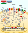
T-cell dysfunction. (A) Tumour-specific CD8+ T-cells need to acquire effector function upon ligation of their TCR with a cognate peptide-MHC complex. Effector function is characterised by expression of cytokines and cytotoxic granules including granzyme B and perforin. Effector T-cells must subsequently apoptosis or, upon appropriate stimulation which promotes metabolic changes, differentiate into long-lived memory cells upon resolution of antigenic challenge. (B) Immunosenescence is characterised by reduced TCR signal transduction, loss of CD28 expression, lower cytokine expression from CD8+ T-cells and a skewed Th1/Th2 ratio in CD4+ T-cells. Fewer naïve T-cells and increases in memory T-cells expressing NK-cell receptors and exhibiting increased innate-like effector functions are also characteristic of senescent T-cells. (C) Senescence is also associated with persistent inflammation. This inflammation also results from chronic infection leading to ‘bystander’ activation of T-cells by inflammatory cytokines. Signalling by cytokines such as IL-6 or TNF-α alter T-cell function, inhibit their differentiation into memory cells and induce apoptosis. (D) Dysregulation and exhaustion of TIL within TME shares some characteristics of senescence and bystander activation. TIL are subject to suppression via VEGF or TGF-β, suffer from metabolic dysregulation due to competition for glucose and amino acids and preventing differentiation into memory T-cells. TIL also express checkpoints due to chronic exposure to antigen. Ligation of these checkpoints with ligands expressed on tumour cells of suppressive immune cells, results in activation of programs of exhaustion based upon action of transcription factors Tox and NR4A blocking effector functions and potentially leading to anergy or apoptosis. Cytotoxic T-lymphocyte-associated protein 4 (CTLA-4), FAS ligand (FASL), Granzyme B (Grb), Interferon-γ (IFN-γ), Interleukin (IL), Nuclear factor kappa B (NF-κB), Programmed cell death protein 1 (PD-1), Programmed cell death protein ligand-1 (PDL-1), Reactive oxygen species (ROS), T-cell immunoglobulin and mucin-domain containing-3 (TIM-3), T-helper cell (Th), TNF-related apoptosis-inducing ligand (TRAIL), Transforming growth factor beta (TGF-β), Tumour necrosis factor-alpha (TNF-α), Vascular endothelial growth factor (VEGF).
Figure 2
Figure 2
Modulation of T-cell function for cancer immunotherapy: T-cell modulation focuses on four aspects of T-cell biology. TCR signalling where inhibition of tyrosine phosphatases such as PTPN22 or SHP1/2 can potentiate T-cell activation and disruption of MAPK signalling may alleviate chronic antigenic stimulation and prevent exhaustion. Co stimulation either with common gamma chain cytokines or agonists of TNSFR receptor signalling may enhance TCR activation and effector function including activation of low affinity T-cells. Cytokines such as IL-7 or IL-21 may also promote proliferation, and subsequently, differentiation into long-lived memory T-cells. Transcriptional regulation of T-cell activation via HDAC-inhibition by butyrate or activation of VDR by 1,25(OH)2D (calcitriol) can regulate T-cell effector function and differentiation. Modulating T-cell metabolism through altering mitochondrial biogenesis to promote effector function or differentiation into memory cells through supplementation with L-arginine or NAD+, or through AMPK activation and mTOR inhibition. Activator protein 1 (AP-1), Adenosine A2a Receptor (ADORA2A), AMP-activated protein kinase (AMPK), Aquaporin-9 (AQP9), Calcineurin (CLN), Carnitine palmitoyltransferase (CPT)-1a, Cbl Proto-Oncogene B (Cbl-b), Cellular Myc (C-Myc), Cytotoxic T-lymphocyte-associated protein 4 (CTLA-4), Dynamin-related protein 1 (Drp1), Eomesodermin (EOMES), Extracellular-signal-regulated kinase (ERK), Fatty acid oxidation (FAO), General control non-depressible 5 (GCN5), Growth Factor Receptor Bound Protein 2 (GRB2), Histone deacetylase (HDAC), Janus associated kinase (JAK), Jun proto-oncogene (C-Jun), Lymphocyte activation gene 3 (LAG-3), Linker for activation of T cells (LAT) lymphocyte-specific protein tyrosine kinase (Lck), Liver kinase B1 (LKB1), Mammalian target of rapamycin (mTOR), Mitogen activated protein kinase (MAPK), Mitogen-activated protein kinase kinase (MEK), Nicotinamide adenine dinucleotide (NADH/NAD+), Nuclear factor kappa-light-chain-enhancer of activated B cells (NF-κB), Nuclear factor of activated T-cells (NFAT), Nuclear Receptor Subfamily 4 Group A Member 1 (NR4A1), Optic atrophy 1 (Opa-1), Oxidative phosphorylation (OXPHOS), Peroxisome proliferator-activated receptor-gamma coactivator-alpha (PGC-1α), phosphoenolpyruvate carboxykinase (PCK1), Phosphoinositide 3-kinases (PI3K), Phosphoinositide pho spholipase C-γ (PLCγ), Programmed cell death protein 1 (PD-1), Protein kinase B (AKT), Protein Tyrosine Phosphatase Non-Receptor Type 22 (PTPN22), Pyruvate Dehydrogenase Kinase 1 (PDK1), Rapidly Accelerated Fibrosarcoma proto-oncogene (RAF), Rat sarcoma virus (RAS), Signal transducer and activator of transcription-5 (STAT5), Sirtuin-1 (Sirt1), Son of sevenless (SOS), Src homology region 2 domain-containing phosphatase (SHP), T cell immunoglobulin and mucin-domain containing-3 (TIM-3), T-box expressed in T cells (T-bet), Thymocyte selection associated high mobility group box (Tox), Triacylglycerol (TAG), Tumour necrosis factor receptor superfamily (TNFRSF), Vitamin D receptor (VDR), Zeta-chain-associated protein kinase-70 (Zap70).
Figure 3
Figure 3
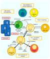
Effective cytotoxic T-cell based immunotherapy requires intervention at different stages: One: nonimmunogenic ‘cold’ tumours, typically expressing little neoantigen or downregulated HLA, will be poorly susceptible to T-cell killing. Immunogenicity of tumour can be improved through use of therapies supporting epitope spread such as radiotherapy or ICD inducing chemotherapy. Two: tumour infiltrating T-cells are subject to induction of tolerance or inhibition through suppressive features of TME or inhibitory immune subsets expressing checkpoints or cytokines such as TGF-β or IL-10 or inhibitory IDO. Three: tumour-specific CD8+ T-cells need to be adequately primed and capable of increased mitochondrial biogenesis to develop into efficacious effector T-cells. Four: in presence of chronic antigenic stimulation effector T-cells can develop an exhausted and dysfunctional phenotype that requires reactivation via ICI. Five: effector T-cells need to be further primed to switch from glycolysis to OXPHOS to generate long lasting anti-tumour CTL.

Similar articles

Cited by

References

    1. Arruga F., Gyau B.B., Iannello A., Vitale N., Vaisitti T., Deaglio S. Immune response dysfunction in chronic lymphocytic leukemia: Dissecting molecular mechanisms and microenvironmental conditions. Int. J. Mol. Sci. 2020;21:1825. doi: 10.3390/ijms21051825. - DOI - PMC - PubMed
    1. Lian J., Yue Y., Yu W., Zhang Y. Immunosenescence: A key player in cancer development. J. Hematol. Oncol. 2020;13:151. doi: 10.1186/s13045-020-00986-z. - DOI - PMC - PubMed
    1. Diao B., Wang C., Tan Y., Chen X., Liu Y., Ning L., Chen L., Li M., Liu Y., Wang G., et al. Reduction and functional exhaustion of T cells in patients with coronavirus disease 2019 (COVID-19) Front. Immunol. 2020;11:827. doi: 10.3389/fimmu.2020.00827. - DOI - PMC - PubMed
    1. Kusnadi A., Ramírez-Suástegui C., Fajardo V., Chee S.J., Meckiff B.J., Simon H., Pelosi E., Seumois G., Ay F., Vijayanand P., et al. Severely ill COVID-19 patients display impaired exhaustion features in SARS-CoV-2-reactive CD8+ T cells. Sci. Immunol. 2021;6:eabe4782. doi: 10.1126/sciimmunol.abe4782. - DOI - PMC - PubMed
    1. Zhang Z., Liu S., Zhang B., Qiao L., Zhang Y., Zhang Y. T cell dysfunction and exhaustion in cancer. Front. Cell Dev. Biol. 2020;8:17. doi: 10.3389/fcell.2020.00017. - DOI - PMC - PubMed