. 2021 Dec 3;9(12):1431.
doi: 10.3390/vaccines9121431.

Longitudinal Characterization of the Mumps-Specific HLA-A2 Restricted T-Cell Response after Mumps Virus Infection

Affiliations

Longitudinal Characterization of the Mumps-Specific HLA-A2 Restricted T-Cell Response after Mumps Virus Infection

Josien Lanfermeijer et al. Vaccines (Basel). .

Abstract

Waning of the mumps virus (MuV)-specific humoral response after vaccination has been suggested as a cause for recent mumps outbreaks in vaccinated young adults, although it cannot explain all cases. Moreover, CD8+ T cells may play an important role in the response against MuV; however, little is known about the characteristics and dynamics of the MuV-specific CD8+ T-cell response after MuV infection. Here, we had the opportunity to follow the CD8+ T-cell response to three recently identified HLA-A2*02:01-restricted MuV-specific epitopes from 1.5 to 36 months post-MuV infection in five previously vaccinated and three unvaccinated individuals. The infection-induced CD8+ T-cell response was dominated by T cells specific for the ALDQTDIRV and LLDSSTTRV epitopes, while the response to the GLMEGQIVSV epitope was subdominant. MuV-specific CD8+ T-cell frequencies in the blood declined between 1.5 and 9 months after infection. This decline was not explained by changes in the expression of inhibitory receptors or homing markers. Despite the ongoing changes in the frequencies and phenotype of MuV-specific CD8+ T cells, TCRβ analyses revealed a stable MuV-specific T-cell repertoire over time. These insights in the maintenance of the cellular response against mumps may provide hallmarks for optimizing vaccination strategies towards a long-term cellular memory response.

Keywords: MMR vaccination; T-cell immunity; mumps infection.


Conflict of interest statement

The authors declare no competing interests.

Figures
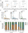
Figure 1
Figure 1
Frequencies of the MuV-specific CD8+ T cells wane after infection. (A) Representative dextramer staining and quantification of the MuV-specific CD8+ T cells against ALD (left), GLM (middle), and LLD (right). (B) Percentages of the MuV-specific CD8+ T cells against ALD (left), GLM (middle), and LLD (right) in HLA-A2-positive individuals upon MuV infection. Solid circles indicate vaccinated individuals, whereas open circles indicate unvaccinated individuals. p.i., post-infection. The percentage of dextramer+ CD8+ T cells of donor 5 have been published before in the study of de Wit et al. 2020 [22]. (C) The relative contribution of the three MuV epitopes to the “total” (=sum of the frequencies of the three) MuV-specific CD8+ T-cell response over time. Donor numbers are depicted above the graphs. Wilcoxon rank test was used to compare T-cell responses of individuals over time.
Figure 2
Figure 2
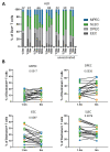
MuV-specific CD8+ T cells differentiate from effector cells towards memory cells over time after infection. (A) Bar graph showing the subset distribution based on CD127 and KLRG-1 expression of the MuV-specific CD8+ T cells against the ALD epitope. Donor numbers are depicted above the graphs. (B) Fraction of the memory subsets based on CD127 and KLRG-1 expression of the MuV-specific CD8+ T cells at 1.5 months and 9 months after MuV infection. The memory precursor cells (MPEC; CD127+, KLRG-1), short-lived effector cells (SLEC; CD127, KLRG-1+), double-positive cells (DPEC; CD127+, KLRG-1+), and the early effector cells (EEC; CD127, KLRG-1). CD8+ T cells specific for the ALD epitope are depicted in orange, for the GLM epitope in blue, and for the LLD epitope in green. Solid circles indicate vaccinated individuals, whereas open circles indicate unvaccinated individuals. Differences between timepoints were tested by Wilcoxon rank test.
Figure 3
Figure 3
MuV-specific T cells tend to home to the bone marrow. (A) The expression of chemokine receptors CCR7 (left), CXCR4 (middle), and CXCR3 (right) based on their MFI value (geometric mean fluorescence intensity) of the MuV-specific T cells at 1.5 months and 9 months after MuV infection. (B) Association between the expression of CCR7 (left) or CXCR4 (right) (geometric mean of MFI) at timepoint 1.5 months post-infection and fold change of dextramer frequencies at timepoints 1.5 months and 9 months after infection. Fold changes were calculated by dividing the expression or frequencies found at 9 months after MuV infection by the expression or frequencies 1.5 months after MuV infection, the calculated fold changes were all below 1, indicating a decrease. Expression of chemokine receptors on CD8+ T cells specific for the ALD epitope are depicted in orange, for the GLM epitope in blue, and for the LLD epitope in green. Solid circles indicate vaccinated individuals, whereas open circles indicate unvaccinated individuals. Differences between timepoints were tested by Wilcoxon rank test. Associations were tested by Spearman’s correlation.
Figure 4
Figure 4
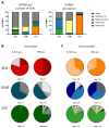
MuV-specific TCRβ repertoire is maintained in the memory phase. (A) Contribution of several Vβ families in the MuV-specific CD8+ T-cell repertoire based on number of sequences (left panel) and based on abundance (right) at 1.5 months of all donors (Table S2A–C). (B,C) Characterization of the T-cell repertoire of MuV-specific CD8+ T cells against ALD, GLM, and LLD, detected by PCR of vaccinated (B) and unvaccinated (C) individuals. Each pie chart depicts the repertoire of a representative donor at a certain timepoint (1.5 months and 9 months after infection). Colors represent shared CDR3 sequences between timepoints and donors. Grey scales depict unique CDR3 sequences. Total number below a pie indicates the number of clones detected. p.i., post-infection.

Similar articles

Cited by

References

    1. MacDonald N., Hatchette T., Elkout L., Sarwal S. Mumps is Back: Why is Mumps Eradication Not Working? Adv. Exp. Med. Biol. 2011;7:197–220. - PubMed
    1. Plotkin S.A., Orenstein W.A., Offit P.A. Vaccines. Elsevier; Philadelphia, PA, USA: 2018.
    1. Kaaijk P., Van Der Zeijst B., Boog M., Hoitink C. Increased mumps incidence in the Netherlands: Review on the possible role of vaccine strain and genotype. Eurosurveillance. 2008;13:18914. doi: 10.2807/ese.13.26.18914-en. - DOI - PubMed
    1. Dayan G.H., Quinlisk M.P., Parker A.A., Barskey A.E., Harris M.L., Schwartz J.M.H., Hunt K., Finley C.G., Leschinsky D.P., O’Keefe A.L., et al. Recent Resurgence of Mumps in the United States. N. Engl. J. Med. 2008;358:1580–1589. doi: 10.1056/NEJMoa0706589. - DOI - PubMed
    1. Karagiannis I., Van Lier A., Van Binnendijk R., Ruijs H., Fanoy E., Spaendonck M.A.E.C.-V., De Melker H., Hahné S. Mumps in a community with low vaccination coverage in the Netherlands. Eurosurveillance. 2008;13:18901. doi: 10.2807/ese.13.24.18901-en. - DOI - PubMed