. 2021 Dec 18;9(12):1499.
doi: 10.3390/vaccines9121499.

Nasal and Salivary Mucosal Humoral Immune Response Elicited by mRNA BNT162b2 COVID-19 Vaccine Compared to SARS-CoV-2 Natural Infection

Affiliations

Nasal and Salivary Mucosal Humoral Immune Response Elicited by mRNA BNT162b2 COVID-19 Vaccine Compared to SARS-CoV-2 Natural Infection

Mariapia Guerrieri et al. Vaccines (Basel). .

Erratum in

Abstract

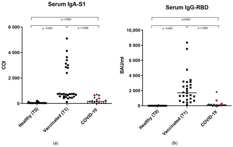
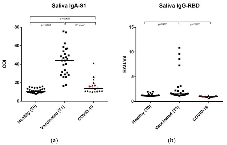
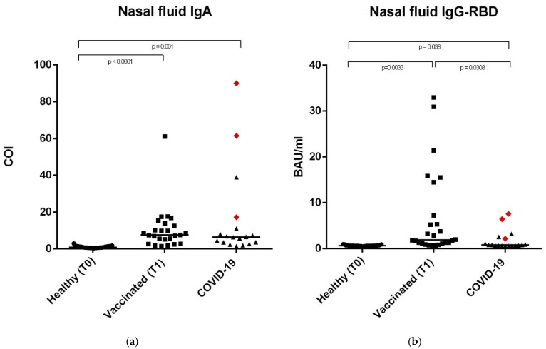
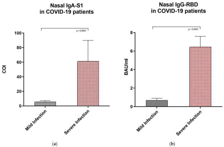
SARS-CoV-2 antibody assays are crucial in managing the COVID-19 pandemic. Approved mRNA COVID-19 vaccines are well known to induce a serum antibody responses against the spike protein and its RBD. Mucosal immunity plays a major role in the fight against COVID-19 directly at the site of virus entry; however, vaccine abilities to elicit mucosal immune responses have not been reported. We detected anti-SARS-CoV-2 IgA-S1 and IgG-RBD in three study populations (healthy controls, vaccinated subjects, and subjects recovered from COVID-19 infection) on serum, saliva, and nasal secretions using two commercial immunoassays (ELISA for IgA-S1 and chemiluminescent assay for IgG-RBD). Our results show that the mRNA BNT162b2 vaccine Comirnaty (Pfizer/BioNTech, New York, NY, USA) determines the production of nasal and salivary IgA-S1 and IgG-RBD against SARS-CoV-2. This mucosal humoral immune response is stronger after the injection of the second vaccine dose compared to subjects recovered from COVID-19. Since there is a lack of validated assays on saliva and nasal secretions, this study shows that our pre-analytical and analytical procedures are consistent with the data. Our findings indicate that the mRNA COVID-19 vaccine elicits antigen-specific nasal and salivary immune responses, and that mucosal antibody assays could be used as candidates for non-invasive monitoring of vaccine-induced protection against viral infection.

Keywords: BNT162b2; COVID-19; IgA; IgG-RBD; SARS-CoV-2; immunity; mucosal; nasal; salivary; vaccine.


Conflict of interest statement

The authors declare no conflict of interest.

Figures

Figure 1
Figure 1
Anti SARS-CoV-2 IgA-S1 and anti SARS-CoV-2 IgG-RBD in serum samples. (a) Serum sample median levels of anti SARS-CoV-2 IgA-S1 in the three study groups expressed as COI (Cut off index). (b) Serum sample median levels of anti SARS-CoV-2 IgG-RBD in the three study groups, expressed as Binding Antibody Units (BAU/mL). In the COVID-19 group, the red rhombuses represent the hospitalized subjects. Statistical analysis and construction of figures were performed with GraphPad Prism 8 Software (GraphPad Software, San Diego, CA, USA). The D’Agostino and Pearson test, Shapiro–Wilk normality test, and Kolmogorov–Smirnov test were used to evaluate non-Gaussian distributions in all study populations. The continuous data were displayed as median and range. Non-parametric results were analyzed with the Mann–Whitney test. For all of the results, p < 0.05 was considered statistically significant.
Figure 2
Figure 2
Anti-SARS-CoV-2 IgA-S1 and anti-SARS-CoV-2 IgG-RBD in saliva samples. (a) Saliva sample median levels of anti-SARS-CoV-2 IgA-S1 in the three study groups, expressed as COI (Cut off index). (b) Saliva sample median levels of anti-SARS-CoV-2 IgG-RBD in the three study groups, expressed as Binding Antibody Units (BAU/mL). In the COVID-19 group, the red rhombuses represent the hospitalized subjects. Statistical analysis and construction of figures were performed with GraphPad Prism 8 Software (GraphPad Software, San Diego, CA, USA). The D’Agostino and Pearson test, the Shapiro-Wilk normality test, and the Kolmogorov–Smirnov test were used to evaluate non-Gaussian distributions in all of the study populations. The continuous data were displayed as median and range. Non-parametric results were analysed with the Mann–Whitney test. For all results, p < 0.05 was considered statistically significant.
Figure 3
Figure 3
Anti SARS-CoV-2 IgA-S1 and anti SARS-CoV-2 IgG-RBD in nasal fluid samples. (a) Nasal fluid sample median levels of anti SARS-CoV-2 IgA-S1 in the three study groups, expressed as COI (Cut off index). (b) Nasal fluid sample median levels of anti SARS-CoV-2 IgG-RBD in the three study groups, expressed as Binding Antibody Units (BAU/mL). In the COVID-19 group, the red rhombuses represent the hospitalized subjects. Statistical analysis and construction of figures were performed with GraphPad Prism 8 Software (GraphPad Software, San Diego, CA, USA). The D’Agostino and Pearson test, Shapiro–Wilk normality test, and Kolmogorov– Smirnov test were used to evaluate the non-Gaussian distributions in all of the study populations. The continuous data were displayed as median and range. Non-parametric results were analysed with the Mann–Whitney test. For all of the results, p < 0.05 was considered statistically significant.
Figure 4
Figure 4
Anti SARS-CoV-2 IgA-S1 and anti SARS-CoV-2 IgG-RBD in nasal fluid samples of subjects recovered from previous mild and severe COVID-19 infection. (a) Nasal secretions median levels of anti SARS-CoV-2 IgA-S1 in subjects with previous mild COVID-19 infection and severe infection, expressed as COI (Cut off index). (b) Nasal fluid samples median levels of anti SARS-CoV-2 IgG-RBD in subjects with previous mild COVID-19 infection and hospitalized subjects, respectively, expressed as Binding Antibody Units (BAU/mL). Statistical analysis and construction of figures were performed with GraphPad Prism 8 Software (GraphPad Software, San Diego, CA, USA). The D’Agostino and Pearson test, Shapiro–Wilk normality test, and Kolmogorov–Smirnov test were used to evaluate non-Gaussian distributions in all study populations. The error bars report the median, and 75° and 25° percentiles. The non-parametric results were analysed with the Mann–Whitney test. For all of the results, p < 0.05 was considered statistically significant.

References

    1. Chao Y.X., Rötzschke O., Tan E.K. The role of IgA in COVID-19. Brain Behav. Immun. 2020;87:182–183. doi: 10.1016/j.bbi.2020.05.057. - DOI - PMC - PubMed
    1. Cervia C., Nilsson J., Zurbuchen Y., Valaperti A., Schreiner J., Wolfensberger A., Raeber M.E., Adamo S., Weigang S., Emmenegger M., et al. Systemic and mucosal antibody responses specific to SARS-CoV-2 during mild versus severe COVID-19. J. Allergy Clin. Immunol. 2021;147:545–557.e9. doi: 10.1016/j.jaci.2020.10.040. - DOI - PMC - PubMed
    1. Pearson C.F., Jeffery R., Thornton E.E., Ahern D.J., Almuttaqi H., Alonzi D.S., Alrubayyi A., Alsaleh G., Bart V.M.T., Batchelor V., et al. Mucosal immune responses in COVID19—A living review. Oxf. Open Immunol. 2021;2:iqab002. doi: 10.1093/oxfimm/iqab002. - DOI - PMC - PubMed
    1. Russell M.W., Moldoveanu Z., Ogra P.L., Mestecky J. Mucosal Immunity in COVID-19: A Neglected but Critical Aspect of SARS-CoV-2 Infection. Front. Immunol. 2020;11:611337. doi: 10.3389/fimmu.2020.611337. - DOI - PMC - PubMed
    1. Fröberg J., Diavatopoulos D.A. Mucosal immunity to severe acute respiratory syndrome coronavirus 2 infection. Curr. Opin. Infect. Dis. 2021;34:181–186. doi: 10.1097/QCO.0000000000000724. - DOI - PubMed

LinkOut - more resources