Review
. 2021 Nov 23;13(12):2344.
doi: 10.3390/v13122344.

Molecular Basis of Epstein-Barr Virus Latency Establishment and Lytic Reactivation

Affiliations
Review

Molecular Basis of Epstein-Barr Virus Latency Establishment and Lytic Reactivation

Takayuki Murata et al. Viruses. .

Abstract

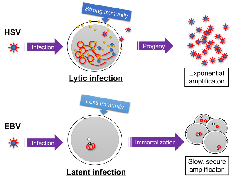
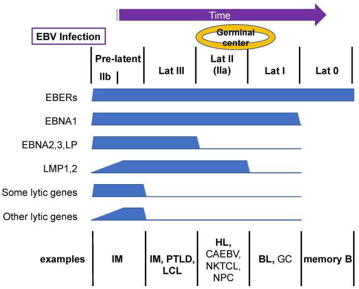
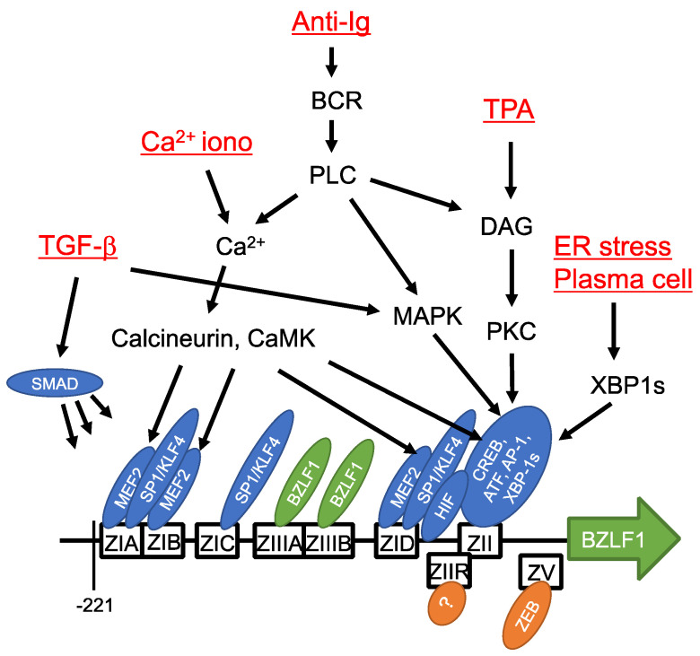
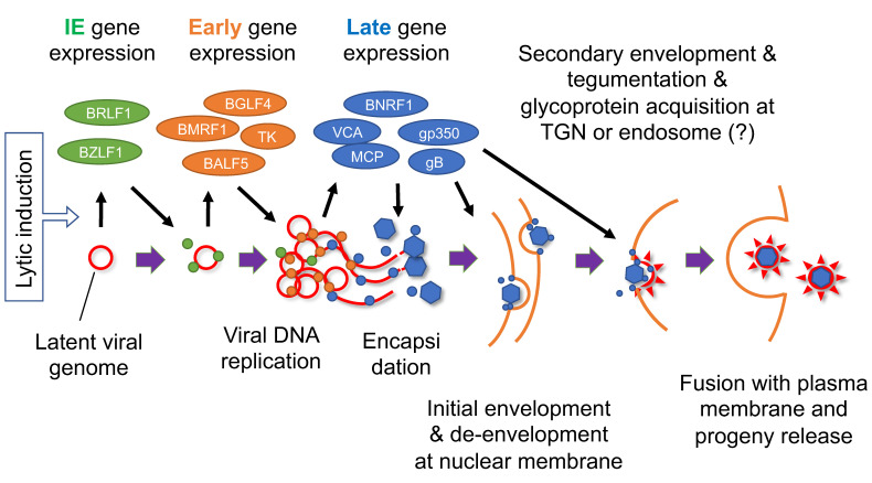
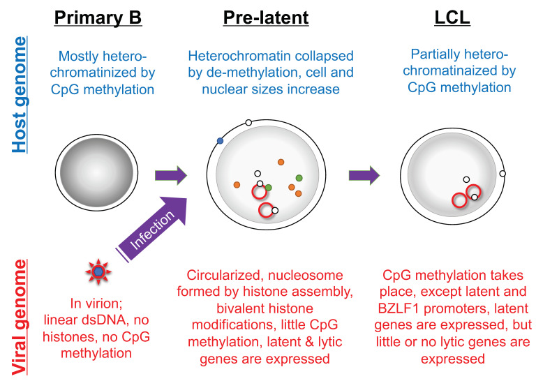
Epstein-Barr virus (EBV) is a causative agent of infectious mononucleosis and several types of cancer. Like other herpesviruses, it establishes an asymptomatic, life-long latent infection, with occasional reactivation and shedding of progeny viruses. During latency, EBV expresses a small number of viral genes, and exists as an episome in the host-cell nucleus. Expression patterns of latency genes are dependent on the cell type, time after infection, and milieu of the cell (e.g., germinal center or peripheral blood). Upon lytic induction, expression of the viral immediate-early genes, BZLF1 and BRLF1, are induced, followed by early gene expression, viral DNA replication, late gene expression, and maturation and egress of progeny virions. Furthermore, EBV reactivation involves more than just progeny production. The EBV life cycle is regulated by signal transduction, transcription factors, promoter sequences, epigenetics, and the 3D structure of the genome. In this article, the molecular basis of EBV latency establishment and reactivation is summarized.

Keywords: EBV; epigenetics; latency; oncogenesis; reactivation; transcription.


Conflict of interest statement

The authors declare no conflict of interest.

Figures

Figure 1
Figure 1
Survival strategies of herpesviruses. Infection of herpes simplex virus (HSV) causes lytic infection and the production of many progeny viruses, although the infected cells and virions may be targeted by the host immune reaction. Latent infection is maintained when the Epstein–Barr virus (EBV) infects a B cell, and the virus genome is amplified in synchronization with host cell amplification after immortalization. Red line indicates viral genome. Yellow and white circles represent viral lytic and latent gene products, respectively.
Figure 2
Figure 2
Transition of EBV gene expression upon primary B-cell infection. EBV infection of primary human B cells triggers the expression of both latent and lytic viral genes, which is termed the pre-latent phase. Subsequently, viral genes are silenced gradually. Example representative cells with the indicated patterns of viral gene expression in vivo are shown. Bold letters are B cells, and non-bold letters indicate non-B cells.
Figure 3
Figure 3
Cis- and trans-acting elements of BZLF1 promoter and signaling pathways. Cis-acting elements of the BZLF1 promoter sequence sufficient for induction in response to lytic stimuli are depicted as boxes. Transcription factors that bind the elements are indicated by ellipses. Inducing reagents/stimuli and signaling molecules are also shown. Modified from [54].
Figure 4
Figure 4
Schematic diagram of EBV lytic cycle. Lytic induction results in a coordinated cascade of viral gene expression and viral DNA replication, followed by encapsidation, envelopment, and progeny production. Red line indicates viral genome. Green, orange, and blue circles represent viral IE, early, and late gene products, respectively. TGN, trans-Golgi network. Modified from [111].
Figure 5
Figure 5
Morphology and epigenetic state of primary B cells infected with EBV. The EBV genome in virions is devoid of nucleosome structure and CpG DNA methylation, but is modified by histones and CpG methylation in infected cells. The host cell morphology and genome are significantly influenced by EBV infection. The red line indicates latent viral genome. Dark gray shadows in nuclei indicate strong heterochromatinization. Green, orange, and blue circles represent viral lytic gene products, and white circles represent viral latent gene products.

References

    1. Young L.S., Yap L.-F., Murray P.G. Epstein–Barr virus: More than 50 years old and still providing surprises. Nat. Rev. Cancer. 2016;16:789–802. doi: 10.1038/nrc.2016.92. - DOI - PubMed
    1. Dunmire S.K., Hogquist K.A., Balfour H.H. Infectious Mononucleosis. Curr. Top. Microbiol. Immunol. 2015;390:211–240. - PMC - PubMed
    1. Cohen J.I. Epstein-Barr virus infection. N. Engl. J. Med. 2000;343:481–492. doi: 10.1056/NEJM200008173430707. - DOI - PubMed
    1. Kenney S.C. Reactivation and lytic replication of EBV. In: Arvin A., Campadelli-Fiume G., Mocarski E., Moore P.S., Roizman B., Whitley R., editors. Human Herpesviruses: Biology, Therapy, and Immunoprophylaxis. Cambridge University Press; Cambridge, UK: 2007. - PubMed
    1. Shannon-Lowe C., Rickinson A. The Global Landscape of EBV-Associated Tumors. Front. Oncol. 2019;9:713. doi: 10.3389/fonc.2019.00713. - DOI - PMC - PubMed

Publication types

MeSH terms

Substances

LinkOut - more resources