. 2021 Nov 28;13(12):2381.
doi: 10.3390/v13122381.

A Novel Picornavirus Discovered in White Leg Shrimp Penaeus vannamei

Affiliations

A Novel Picornavirus Discovered in White Leg Shrimp Penaeus vannamei

Shuang Liu et al. Viruses. .

Abstract

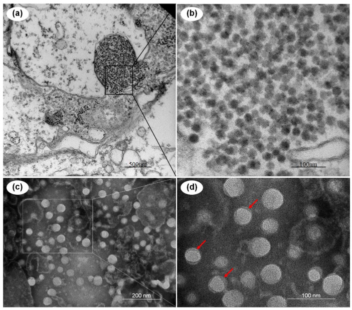
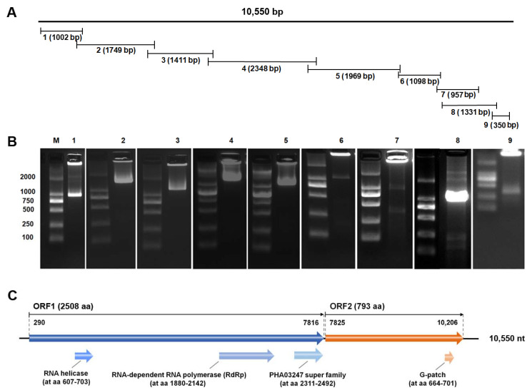
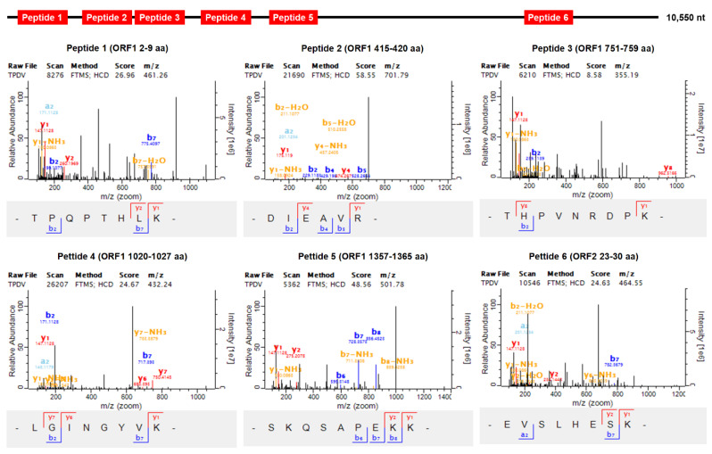
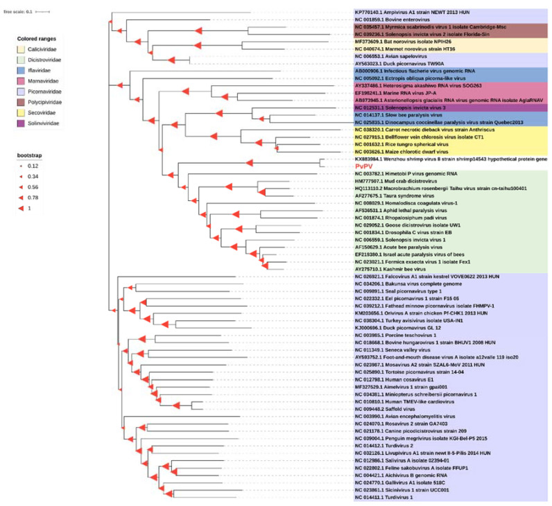
Global shrimp farming is increasingly threatened by various emerging viruses. In the present study, a novel picornavirus, Penaeus vannamei picornavirus (PvPV), was discovered in moribund White leg shrimp (Penaeus vannamei) collected from farm ponds in China in 2015. Similar to most picornaviruses, PvPV is non-enveloped RNA virus, with a particle diameter of approximately 30 nm. The sequence of the positive single-stranded RNA genome with a length of 10,550 nts was characterized by using genome sequencing and reverse transcription PCR. The existence of PvPV related proteins was further proved by confirmation of viral amino acid sequences, using mass spectrometry analysis. Phylogenetic analysis based on the full-length genomic sequence revealed that PvPV was more closely related to the Wenzhou shrimp virus 8 than to any other dicistroviruses in the order Picornavirales. Genomic sequence conservative domain prediction analysis showed that the PvPV genome encoded a large tegument protein UL36, which was unique among the known dicistroviruses and different from other dicistroviruses. According to these molecular features, we proposed that PvPV is a new species in the family Dicistroviridae. This study reported the first whole-genome sequence of a novel and distinct picornavirus in crustaceans, PvPV, and suggests that further studies of PvPV would be helpful in understanding its evolution and potential pathogenicity, as well as in developing diagnostic techniques.

Keywords: Dicistroviridae; Penaeus vannamei; Penaeus vannamei picorna viruses (PvPV); Picornavirales; shrimp.


Conflict of interest statement

The authors declare no conflict of interest.

Figures

Figure 1
Figure 1
Transmission electron micrographs of the Penaeus vannamei picornavirus (PvPV) virions and an ultrathin section of the hepatopancreatic epithelial cells of the moribund Penaeus vannamei from the farm in Shandong, China. (a,b) Ultrathin section of hepatopancreatic epithelial cells; (c,d) purified PvPV virions; (bd) show magnified micrographs in the corresponding framed areas of (ac), respectively. Note the tegument of PvPV particles, indicated with red arrows. Scale bars: (a) 500 nm, (b) 100 nm, (c) 200 nm, (d) 100 nm.
Figure 2
Figure 2
Assembly strategy and agarose gel electrophoresis of reverse transcription PCR (RT-PCR) products of Penaeus vannamei picornavirus (PvPV) genome sequence, and schematic presentation of the genomic open reading frame segments and conserved domains of PvPV. (A) Strategy of re-cloning and assembling the genomic open reading frame of PvPV by RT-PCR. The numbers 1–9 corresponding to lanes 1–9 in Figure (B) represent the expected size of the RT-PCR amplification products and indicate the position of amplicons in the PvPV genome. (B) Agarose gel electrophoresis of RT-PCR products for re-cloning of the PvPV genomic sequence. Lane M: 2000 bp marker; lanes 1–9: amplicons of PvPV genomic sequence. (C) The potential protein encoding by segments of PvPV genome was identified by using open reading frame (ORF), and the conserved protein domains were predicted using the conserved domain database (CDD) searching tool from the National Center for Biotechnology Information website.
Figure 3
Figure 3
Mass spectrometry analysis of Penaeus vannamei picornavirus (PvPV) proteins. To identify PvPV protein by matrix-assisted laser desorption ionization time-of-flight mass spectrometry analysis, six peptides were screened and measured in the digested PvPV protein using trypsin.
Figure 4
Figure 4
Phylogeny analysis of the Penaeus vannamei picornavirus (PvPV) and other picornaviruses. Phylogenetic trees based on the full-length genomic sequence were constructed using the neighbor-joining method with 1000 bootstrap replicates under the parameter of complete deletion and Poisson model using the software MEGA 6.0. The size of the red triangle symbol at the branch nodes represents the bootstrap confidence levels of the 1000 bootstrap replications obtained. Scale bar was 0.1. The PvPV is highlighted in bold red.

References

    1. Flegel T.W., Lightner D.V., Lo C.F., Owens L. Shrimp Disease Control: Past, Present and Future. Dis. Asian Aquac. VI. Fish Health Sect. Asian Fish. Soc. Manila Philipp. 2008;505:355–378.
    1. Flegel T.W. Historic emergence, impact and current status of shrimp pathogens in Asia. J. Invertebr. Pathol. 2012;110:166–173. doi: 10.1016/j.jip.2012.03.004. - DOI - PubMed
    1. Soto M.A., Lotz J.M. Epidemiological Parameters of White Spot Syndrome Virus Infections in Litopenaeus vannamei and L. setiferus. J. Invertebr. Pathol. 2001;78:9–15. doi: 10.1006/jipa.2001.5035. - DOI - PubMed
    1. Soto M.A., Shervette V.R., Lotz J.M. Transmission of white spot syndrome virus (WSSV) to Litopenaeus vannamei from infected cephalothorax, abdomen, or whole shrimp cadaver. Dis. Aquat. Org. 2001;45:81–87. doi: 10.3354/dao045081. - DOI - PubMed
    1. Zhang Q., Liu Q., Liu S., Yang H., Liu S., Zhu L., Yang B., Jin J., Ding L., Wang X., et al. A new nodavirus is associated with covert mortality disease of shrimp. J. Gen. Virol. 2014;95:2700–2709. doi: 10.1099/vir.0.070078-0. - DOI - PubMed

Publication types

LinkOut - more resources