Review
. 2021 Dec 4;13(12):2435.
doi: 10.3390/v13122435.

A Review of the Emerging White Chick Hatchery Disease

Affiliations
Review

A Review of the Emerging White Chick Hatchery Disease

Kerry McIlwaine et al. Viruses. .

Abstract

White chick hatchery disease is an emerging disease of broiler chicks with which the virus, chicken astrovirus, has been associated. Adult birds typically show no obvious clinical signs of infection, although some broiler breeder flocks have experienced slight egg drops. Substantial decreases in hatching are experienced over a two-week period, with an increase in mid-to-late embryo deaths, chicks too weak to hatch and pale, runted chicks with high mortality. Chicken astrovirus is an enteric virus, and strains are typically transmitted horizontally within flocks via the faecal-oral route; however, dead-in-shell embryos and weak, pale hatchlings indicate vertical transmission of the strains associated with white chick hatchery disease. Hatch levels are typically restored after two weeks when seroconversion of the hens to chicken astrovirus has occurred. Currently, there are no commercial vaccines available for the virus; therefore, the only means of protection is by good levels of biosecurity. This review aims to outline the current understanding regarding white chick hatchery disease in broiler chick flocks suffering from severe early mortality and increased embryo death in countries worldwide.

Keywords: chicken astrovirus; hatchery disease; vertical transmission; white chick.


Conflict of interest statement

The authors declare no conflict of interest.

Figures

Figure 1
Figure 1
A schematic diagram of the poultry breeding pyramid illustrating the typical timeline of white chick hatchery disease during parent stock egg-laying (yellow line). The dip in the yellow line indicates a typical CAstV infection and subsequent hatchery drop with vertical transmission of virus, resulting in white chick hatchery disease in broiler progeny prior to parent flock seroconversion against CAstV and consequent recovery of hatch levels.
Figure 2
Figure 2
Phylogenetic tree of CAstV complete ORF 2 amino acid sequences. Sequences clustered according to sub(geno)groupings of the A (subgroups Ai, Aii and Aiii) and B (subgroups Bi, Bii, Biii and Biv) groups, which are serologically distinct. The maximum likelihood tree was constructed in RAxML version 8 [51] on Geneious version 2021.1 (Biomatters) using the GAMMA BLOSUM62 protein model with 1000 rapid bootstrapping replicates and searching for best-scoring ML tree with 456 parsimony random seed.
Figure 3
Figure 3
Multiple sequence alignment of representative B-group CAstV capsid protein surface spike domains. The Bi-group surface spike domain sequence was from strain JN582327_11672, the Bii-group from strain JN582316_VF08-3, the Biii-group from strain KC618325_PDRC/1803/South and the B iv-group from strain MT789787_19-0981/19_Canada *. Conserved, similar and non-conserved amino acids are highlighted in dark blue, light blue and white, respectively. Two clusters of amino acid residues that are candidates to form putative avian cell receptor-binding sites (annotated as ‘Site A’ and ‘Site B’) of the spike domain are boxed in red. Site A consists of residues 532–540 (532–542 for the Bii-group CAstV) and site B consists of residues 552–561 (554–563 for the B ii-group CAstV). Site B contains a highly conserved glutamine residue, indicated by an asterisk, that is the structural equivalent to Q552 of the turkey TAstV-2 capsid protein spike domain, and deletion of which results in increased incidence of disease in turkeys infected with TAstV-2 [59,60]. Alignments were performed with the online ClustalW server [61].
Figure 4
Figure 4
Comparative protein modelling reveals structural differences between CAstV capsid surface spike domains of the B-groups of the virus. Each cartoon model represents a side view of one dimer subunit of the CAstV capsid surface spike domain with the N-terminal located at the bottom and the homodimer interface on the right-hand side. (a) Bi-type spike domain from strain JN582327_11672; (b) Bii-type spike domain from strain JN582316_VF08-3; (c) Biii-type spike domain from strain KC618325_PDRC/1803/South; and (d) Biv-type spike domain from strain MT789787_19-0981/19_Canada*. The residues modelled are indicated in parentheses. Although the modelled proteins share the same general α/β structure, consisting of a scaffold that comprises an antiparallel β-barrel with a tightly packed hydrophobic core, differences that define each of the B-type spike domains are apparent. The homology models were built and refined with MODELLER [62] using the 1.5-Å resolution crystal structure of turkey astrovirus 2 (TAstV-2) capsid surface spike domain (PDB ID: 3TS3) as the structural template [59]. The models were visualised using the PyMOL Molecular Graphics System [63].
Figure 5
Figure 5
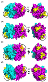
Identification of putative avian receptor-binding sites in B-group CAstV capsid protein spike domains. Structural models of the CAstV B-group spike domains are shown in the biologically relevant homodimer form and depicted in side-view as surface representations. The dimers were built by mapping the homology model monomers described in Figure 4 onto the coordinates of the TAstV-2 spike domain homodimer [59]. One dimer subunit is coloured cyan and the other is coloured magenta. Amino acid residues suggested by sequence alignment (Figure 3) to form potential avian cell receptor-binding sites are coloured yellow. These form two distinct surface-exposed clusters circled in black and annotated as Site A and Site B, located diagonally opposite one another on the top and bottom edges of each dimer subunit. In each model, a highly conserved glutamine residue (located in Site B) that is the structural equivalent of Q552 of the turkey astrovirus TAstV-2 spike domain is coloured orange. We speculate that amino acid differences between the putative receptor-binding site regions in the different B-group viruses could form the molecular basis for receptor recognition and binding and could also explain differences in disease incidence of the B-group CAstVs. It is intriguing to further speculate that each B-group CAstV may recognise and bind at least one different cell receptor. (a) Bi–group spike domain homodimer from strain JN582327_11672; (b) Bii–group spike domain homodimer from strain JN582316_VF08-3; (c) Biii–group spike domain homodimer from strain KC618325_PDRC/1803/South; and (d) Biv–group spike domain homodimer from strain MT789787_19-0981/19_Canada*. The models were visualised using the PyMOL Molecular Graphics System [63].

References

    1. Mazaheri A., Prusas C., Voß M., Hess M. Vertical transmission of fowl adenovirus serotype 4 investigated in specified patho-gen-free birds after experimental infection. Arch. Fur Geflugelkd. 2003;67:6–10.
    1. Todd D., Wilkinson D.S., Jewhurst H.L., Wylie M., Gordon A.W., Adair B.M. A seroprevalence investigation of chicken astrovirus infections. Avian Pathol. 2009;38:301–309. doi: 10.1080/03079450903055421. - DOI - PubMed
    1. Linden J. Vertically Transmitted Health Issues in Poultry. [(accessed on 6 May 2021)]. Available online: https://thepoultrysite.com/articles/vertically-transmitted-health-issues...
    1. Smyth V., Trudgett J., Wylie M., Jewhurst H., Conway B., Welsh M., Kaukonen E., Perko-Mäkelä P. Chicken astrovirus detected in hatchability problems associated with ‘white chicks’. Vet. Rec. 2013;173:403–404. doi: 10.1136/vr.f6393. - DOI - PubMed
    1. Long K.E., Hastie G.M., Ojkić D., Brash M.L. Economic impacts of white chick syndrome in Ontario, Canada. Avian Dis. 2017;61:402–408. doi: 10.1637/11592-012217-CaseR. - DOI - PubMed

Publication types

MeSH terms

LinkOut - more resources