Review
. 2021 Dec 9;13(12):2465.
doi: 10.3390/v13122465.

The Nucleocapsid of Paramyxoviruses: Structure and Function of an Encapsidated Template

Affiliations
Review

The Nucleocapsid of Paramyxoviruses: Structure and Function of an Encapsidated Template

Louis-Marie Bloyet. Viruses. .

Abstract

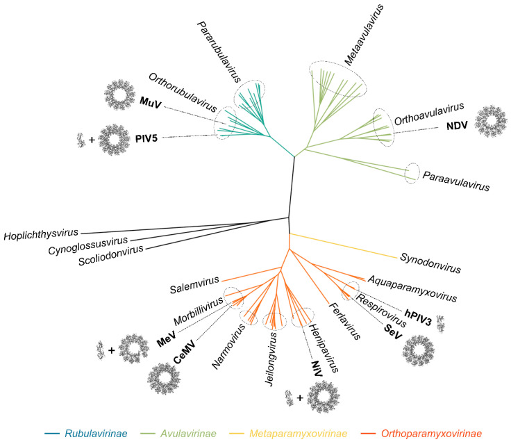
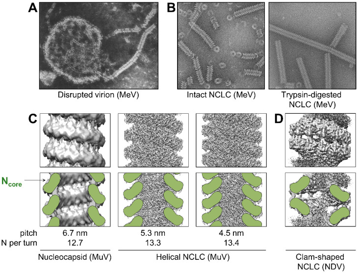
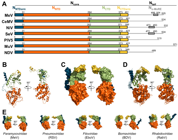
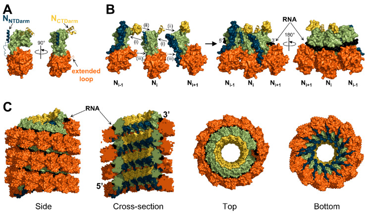
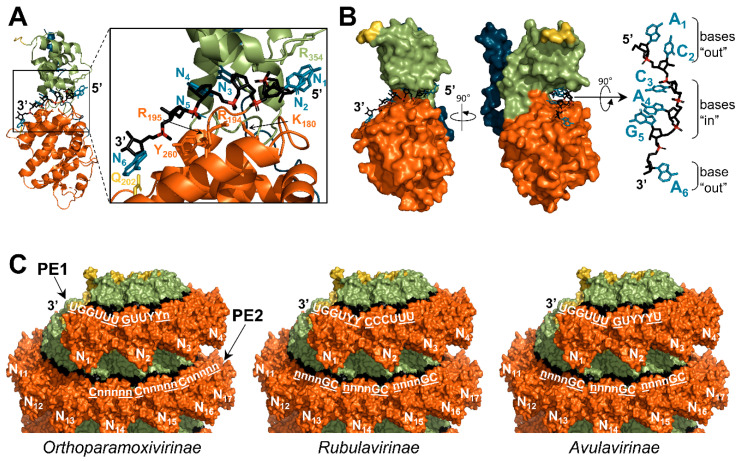
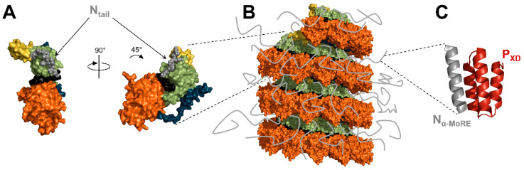
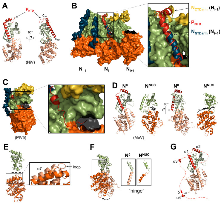
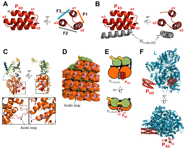
Viruses of the Paramyxoviridae family share a common and complex molecular machinery for transcribing and replicating their genomes. Their non-segmented, negative-strand RNA genome is encased in a tight homopolymer of viral nucleoproteins (N). This ribonucleoprotein complex, termed a nucleocapsid, is the template of the viral polymerase complex made of the large protein (L) and its co-factor, the phosphoprotein (P). This review summarizes the current knowledge on several aspects of paramyxovirus transcription and replication, including structural and functional data on (1) the architecture of the nucleocapsid (structure of the nucleoprotein, interprotomer contacts, interaction with RNA, and organization of the disordered C-terminal tail of N), (2) the encapsidation of the genomic RNAs (structure of the nucleoprotein in complex with its chaperon P and kinetics of RNA encapsidation in vitro), and (3) the use of the nucleocapsid as a template for the polymerase complex (release of the encased RNA and interaction network allowing the progress of the polymerase complex). Finally, this review presents models of paramyxovirus transcription and replication.

Keywords: intrinsic disorder; nucleocapsid; nucleoprotein; paramyxoviruses; phosphoprotein; polymerase complex.


Conflict of interest statement

The author declares no conflict of interest.

Figures

Figure 1
Figure 1
Phylogenetic tree of the Paramyxoviridae family. The phylogenetic tree was generated from an alignment of full-length L proteins. One sequence per species was used. The 78 sequences were selected based on the GenBank accession numbers given by the 2020 taxonomy of the International Committee on Taxonomy of Viruses. The names of the genera are indicated in italics. In bold, viruses for which structural data on the N protein is available. Structures of rings or single helical turns of the nucleocapsid-like complexes are shown from a top view (Newcastle disease virus (NDV), reconstructed from PDB: 6JC3; Sendai virus (SeV), reconstructed from PDB: 6M7D; Nipah virus (NiV), PDB: 7NT5; cetacean morbillivirus (CeMV), PDB: 7OI3; measles virus (MeV), PDB: 6h5Q; parainfluenza virus 5 (PIV5), PDB: 4XJN; mumps virus (MuV), PDB: 7EWQ). For human parainfluenza 3 (hPIV3), PIV5, MeV, and NiV, the structures of the N0-P complexes are shown (hPIV3, PDB: 7EV8; PIV5, PDB: 5WKN; MeV, PDB: 5E4V; NiV, PDB: 4CO6).
Figure 2
Figure 2
Organization and structure of the components of the RNA synthesis machinery. (A) Schematic representation of the viral genome and the regulatory elements. The genome contains at least six conserved adjacent genes separated by intergenic regions (IR). These genes encode for the nucleoprotein (N), the phosphoprotein (P), the matrix protein (M), the fusion protein (F), the receptor-binding protein (RBP), and the large protein (L). The promoters located at the 3′ end of the genome (transcription and replication) and the antigenome (replication only) are bipartite and made of two promoter elements (PE1 and PE2). The transcription of each gene starts on a “gene start” signal (GS, in yellow) and ends on a “gene end” signal (GE, orange). (B) Cartoon representation of the structure of the protein components of the RNA synthesis machinery of PIV5 (N, PDB: 4XJN; PNTD, PDB: 5WKN; POD, PXD, and L, PDB: 6V85). Disordered regions are represented as dotted lines. (C) Schematic representation of viral transcription and replication. Encapsidated genomes are transcribed into a gradient of mRNAs. Replication of the genomes requires the production of encapsidated antigenome intermediates.
Figure 3
Figure 3
Conformations of nucleocapsids. (A) Disrupted measles particle by negative-stain microscopy. Republished with permission of Journal of Virology, from [116]; permission conveyed through Copyright Clearance Center, Inc. (B) Intact (left) and trypsin-digested (right) nucleocapsid-like complexes (NCLC) of MeV. Republished with permission of Journal of Virology, from [117]; permission conveyed through Copyright Clearance Center, Inc. (C) Top row: density maps of MuV nucleocapsid (left, EMDB: EMD-2630) and MuV NCLC (center, EMBD: EMD-31368; right, EMBD: EMD-31369). Bottom row: cross sections of the density maps shown above with schematic representation of Ncore in green. The pitch and number of N protomers per helical turn are indicated at the bottom. (D) Top: density map of a clam-shaped structure of NDV (EMBD: EMD-9793). Bottom: cross section of the density map shown above with schematic representation of Ncore in green.
Figure 4
Figure 4
Organization and structure of the nucleoproteins. (A) Organization and boundaries of the domains of all nucleoproteins whose structure has been solved. Boundaries of Nα-MoRE are only known for MeV [71,73], NiV [74,127], and SeV [128]. (B) Structure of the N protein of MeV in cartoon representations (PDB: 6H5Q). (C) Structure of the N protein of MeV in surface representations (PDB: 6H5Q). (D) Superimposition of aligned structures of MeV (PDB: 6H5Q), CeMV (PDB: 7OI3), NiV (PDB: 7NT5), SeV (PDB: 6M7D), PIV5 (PDB: 4XJN), MuV (PDB: 7EWQ), and NDV (PDB: 6JC3). (E) Surface representations of the structure of the N proteins of MeV (PDB: 6H5Q), respiratory syncytial virus (RSV, PDB: 2WJ8), Ebola virus (EboV, PDB: 5Z9W), Borna disease virus (BDV, PDB: 1N93), and rabies virus (RabV, PDB: 2GTT). All the structures shown in Figure 4 correspond to N proteins observed in their assembled form (i.e., in NCLC).
Figure 5
Figure 5
Mode of assembly. (A) Structure of the nucleoprotein of PIV5 shown in surface representation, except for the NNTDarm, NCTDarm, and extended loop, which are shown in cartoon representation (PDB: 4XJN). (B) Surface representation of three N protomers with the NNTDarm, NCTDarm, and extended loop of the Ni protomer shown in cartoon representation. The RNA is shown in black. (C) Surface representation of a nucleocapsid-like complex of MeV shown with four different views (reconstituted from PDB: 6H5Q). Color code is the same as in Figure 4.
Figure 6
Figure 6
Position of the RNA. (A) Cartoon representation of MeV nucleoprotein bound to the first six nucleotides of the genome (5′ end) (PDB: 6H5S). The RNA and the residues implicated in the binding to the RNA are represented with sticks. The bases of the RNA are shown in blue, the backbone in black, and the phosphates in red. Residue Q202 is shown in yellow. (B) Same as (A) with the nucleoprotein in surface representation. (C) NCLCs of CeMV are shown in surface representation (reconstitution of the helix from PDB: 7OI3). For each subfamily, the consensus sequences of the promoter elements (PE1 and PE2) are shown in white. Well-conserved nucleotides are in capital letters. Bases in “out” positions are underlined. For N, same color code as in Figure 4.
Figure 7
Figure 7
Structure and position of Ntail. (A) Structure of the nucleoprotein of SeV in surface representation with the residues of Ntail shown with spheres (PDB: 6M7D). (B) Surface representation of the nucleocapsid-like complex of CeMV with the Ntail represented as grey lines (reconstitution from PDB: 7OI3). (C) Structure of MeV PXD in complex with Nα-MoRE (PDB: 1T6O). For N, the color code is the same as in Figure 4.
Figure 8
Figure 8
Structure of the N0-P complex. (A) Structure of the N0-P complex of NiV (PDB: 4CO6). The NNTD and the NCTD are in pale orange and pale green, respectively. (B) Superimposition of the structure of three N protomers of NiV NLPC (PDB: 7NT5) with NiV N0-P (PDB: 4CO6). The NCTD of the N0-P complex was aligned onto the NCTD of the Ni promoter. The N protomers are presented in surface representation, while the NNTDarm, NCTDarm, and the extended loop of the Ni protomer are shown in cartoon representation. PNTD is shown in cartoon representation. (C) Structure of the N0-P complex of PIV5 with PNTD in cartoon representation (PDB: 5WKN). The RNA is shown as a transparent black surface. (D) Side-by-side comparison of the structures of the N0-P complex (PDB: 5E4V) and of an N protomer of the NCLC of MeV (PDB: 6H5Q) (NNTDarm and NCTDarm are not shown). The NNTD and the NCTD of N0 are in pale orange and pale green, respectively. (E) Superimposition of the NCTD (top) and of the NNTD (bottom) of the N0-P complex and of an N protomer of the NCLC of MeV. (F) Superimposition of the structures of MeV N0 and NNUC (NNTDarm and NCTDarm are not shown). The structures are aligned based on their NCTD. (G) Structure of the MeV N0-P complex with the disordered region downstream PNTD shown as a dotted line. The transient alpha-helices (α3 and α4) are shown in cartoon representation.
Figure 9
Figure 9
Interaction network of PXD. (A) Structure of MeV PXD (PDB: 1T6O). The three faces of the “prism” are indicated (F1, F2, F3). F1, F2, and F3 are shown with a color code corresponding to the color used to draw the protein partner. (B) Structure of MeV PXD in complex with Nα-MoRE (PDB: 1T6O). (C) Structure of a protomer of MeV N, as observed in the NCLC (PDB: 6H5Q) with the RNA shown in black. Amino acids shown as red spheres correspond to CDV N residues that once mutated restore the growth of a recombinant CDV bearing deletions in the NCDR [191]. The amino acids corresponding to the residues of NiV identified as important for the interaction with PXD are shown as pink spheres [192]. Bottom: enlargements of the NNTD with the acidic loop shown in pink. (D) Surface representation of a nucleocapsid-like complex of MeV (reconstituted from PDB: 6H5Q) with the acidic loop shown in pink. (E) Schematic representation of two adjacent N protomers bound to PXD in complex with Nα-MoRE. (F) Structure of the polymerase complex of PIV5 (PDB: 6V85). For panels (CE), N protomers are colored according to the color code in Figure 4.
Figure 10
Figure 10
Summary of experimental data on the effect of Ntail on the polymerase’s activity. Schematic representation of recombinant N proteins analyzed in minigenome assays. The levels of gene expression are normalized to the level of the wild-type protein (first protein of each set). Activity levels: 0–10%: “-”; 50–75%: “+++”; 75–100%: “++++”; >100%: “+++++”. For mutants (6) and (7) Nα-MoRE is inserted in a loop of NNTD at residue 138. Mutants (4), (5), (6), (9), and (11) contain internal deletion shown with a “^”. Activity levels are inferred from the references indicated on the right.
Figure 11
Figure 11
Models of different steps of RNA synthesis. Schematic representations of the recruitment of the polymerase complex (A), the initiation of RNA synthesis (B), the release of the RNA (C), the dynamics of the interaction between PXD and Ntail and of PXD-Nα-MoRE with Ncore (DK), and the encapsidation of nascent RNA during replication (LN). (A) The polymerase complex is recruited on its template via the interaction between PXD and Ntail. (B) The polymerase complex finds the 3′ end of the genome to initiate RNA synthesis. Bottom panel: enlargement of the top panel. The binding of PNTD to the first protomer may enhance the release of the RNA and its engagement in the template entrance channel. (C) The progression of the polymerase complex induces a local rearrangement of the helix α7 and the preceding loop. This conformational change releases the RNA. (DK) Two models are proposed for the dynamics of the interaction between PXD and N during the progression of the polymerase complex. (D,H) In both cases, the polymerase induces a local conformational change on the helical nucleocapsid. The Ntail are flexible, disordered, and inhibit the progression of the polymerase complex. (E,I) PXD binds Ntail and induces the folding of the Nα-MoRE. (F) In model 1, the PXD/Nα-MoRE complex binds the Ncore of the next N protomer. (G) The stabilization of Ntail by PXD allows the polymerase complex to continue RNA synthesis. (J) In model 2, the PXD/Nα-MoRE complex binds the Ncore of an N protomer from the previous helical turn. (K) The anchoring of the Ntail to the previous helical turn “clears the way” and allows an efficient progression of the polymerase complex. (L) During replication, some PNTD are bound to N0 monomers. The N0-P complex is stabilized by the transient α4 and the preceding delta domain. N0 has a strong affinity for the first six residues of the viral genome. (M) N0 is transferred onto the nascent RNA and switches from the N0 to the NNUC conformation. (N) Thanks to interactions between the N0 and the last N protomer added on the nascent RNA, the N proteins can be added to the growing polymerase even if the affinity for the RNA sequence is low. For N, the color code is the same as in Figure 4.

References

    1. Rima B., Balkema-Buschmann A., Dundon W.G., Duprex P., Easton A., Fouchier R., Kurath G., Lamb R., Lee B., Rota P., et al. ICTV Virus Taxonomy Profile: Paramyxoviridae. J. Gen. Virol. 2019;100:1593–1594. doi: 10.1099/jgv.0.001328. - DOI - PMC - PubMed
    1. Cox R., Plemper R.K. The paramyxovirus polymerase complex as a target for next-generation anti-paramyxovirus therapeutics. Front. Microbiol. 2015;6:459. doi: 10.3389/fmicb.2015.00459. - DOI - PMC - PubMed
    1. Aggarwal M., Plemper R.K. Structural Insight into Paramyxovirus and Pneumovirus Entry Inhibition. Viruses. 2020;12:342. doi: 10.3390/v12030342. - DOI - PMC - PubMed
    1. Emerson S.U., Wagner R.R. Dissociation and reconstitution of the transcriptase and template activities of vesicular stomatitis B and T virions. J. Virol. 1972;10:297–309. doi: 10.1128/jvi.10.2.297-309.1972. - DOI - PMC - PubMed
    1. Emerson S.U., Yu Y. Both NS and L proteins are required for in vitro RNA synthesis by vesicular stomatitis virus. J. Virol. 1975;15:1348–1356. doi: 10.1128/jvi.15.6.1348-1356.1975. - DOI - PMC - PubMed

Publication types

LinkOut - more resources