Review
. 2021 Dec;37(6):395-424.
doi: 10.3393/ac.2021.00927.0132. Epub 2021 Dec 22.

Surgical Treatment of Low-Lying Rectal Cancer: Updates

Affiliations
Review

Surgical Treatment of Low-Lying Rectal Cancer: Updates

Cristopher Varela et al. Ann Coloproctol. 2021 Dec.

Abstract

Despite innovative advancements, distally located rectal cancer remains a critical disease of challenging management. The crucial location of the tumor predisposes it to a circumferential resection margin (CRM) that tends to involve the anal sphincter complex and surrounding organs, with a high incidence of delayed anastomotic complications and the risk of the pelvic sidewall or rarely inguinal lymph node metastases. In this regard, colorectal surgeons should be aware of other issues beyond total mesorectal excision (TME) performance. For decades, the concept of extralevator abdominoperineal resection to avoid compromised CRM has been introduced. However, the complexity of deep pelvic dissection with poor visualization in low-lying rectal cancer has led to transanal TME. In contrast, neoadjuvant chemoradiotherapy (NCRT) has allowed for the execution of more sphincter-saving procedures without oncologic compromise. Significant tumor regression after NCRT and complete pathologic response also permit applying the watch-and-wait protocol in some cases, now with more solid evidence. This review article will introduce the current surgical treatment options, their indication and technical details, and recent oncologic and functional outcomes. Lastly, the novel characteristics of distal rectal cancer, such as pelvic sidewall and inguinal lymph node metastases, will be discussed along with its tailored and individualized treatment approach.

Keywords: Lymph node excision; Proctectomy; Rectal neoplasms; Surgical procedures.


Conflict of interest statement

No potential conflict of interest relevant to this article was reported.

Figures
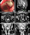
Fig. 1.
Fig. 1.
(A) Colonoscopy finding of low-lying ulcerofungating mass at the anterolateral part of the distal rectum. (B) Rectal magnetic resonance imaging showing mass abutting the prostate gland. (C) Axial imaging of negative circumferential resection margin at prostate level. (D) Tumor extension to the left levator ani muscle. (E) Unilateral tumor involvement of pelvic floor. (F) Enlarged pelvic lymph nodes at the left pelvic sidewall.
Fig. 2.
Fig. 2.
(A) After neoadjuvant chemoradiotherapy, colonoscopy imaging findings showed marked shrinkage in mass. (B) Rectal magnetic resonance imaging (MRI) showed downstaging for cT and cN. (C) Axial rectal MRI shows tumor abutting to left levator ani muscle. (D) Coronal rectal MRI shows downsizing of left pelvic sidewall lymph node (6.3 mm).
Fig. 3.
Fig. 3.
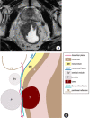
(A) Tumor abutting the prostate capsule (left) and surgical planes of anterior pelvic dissection with customized excision of Denovilliers fascia at the prostate level. (B) Schematic representation of anterior planes of dissection. Adapted from Kim et al. [1] according to the Creative Commons License.
Fig. 4.
Fig. 4.
The tumor center is located anterolaterally at the prostate gland, extending to the right levator ani muscle.
Fig. 5.
Fig. 5.
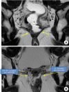
(A) Positron emission tomography showed fluorodeoxyglucose high uptake at the right pelvic sidewall. (B) Rectal magnetic resonance imaging also showed a metastatic pelvic lymph node at a right pelvic wall.
Fig. 6.
Fig. 6.
Rectal magnetic resonance imaging (MRI) showed mesorectal metastatic lymph node (LN) (A) and right metastatic LN at the pelvic wall (B). After chemoradiotherapy, rectal MRI showed a decrease in the right internal iliac LN in size; from 7.3 to 4.9 mm, from 4.5 mm to 4.2 mm respectively, still metastatic LN (C). After robotic partial excision of levator ani muscle and right pelvic LN dissection, a remaining right pelvic LN was present in the follow-up rectal MRI 3 months after surgery (D).
Fig. 7.
Fig. 7.
(A) After specimen resection, the distal portion of the seminal vesicles is observed covered by Denovilliers fascia (DVF), but the prostate capsule is exposed after DVF partial excision at the prostate level. (B) Sagittal rectal magnetic resonance imaging showed the level of the tumor.
Fig. 8.
Fig. 8.
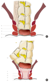
Surgical strategies for rectal resection. LAM, levator ani muscle; DL, dentate line; IAS, internal anal sphincter; EAS, external anal sphincter; ISS, intersphincteric space; ISG, intersphincteric groove; uLAR, ultralow rectal resection; ISR, intersphincteric resection; HLE, hemilevator excision; PELM, partial excision of LAM; APR, abdominoperineal resection; ELAPE, extralevator abdominoperineal excision.
Fig. 9.
Fig. 9.
An ultralow anterior resection. (B) After ultralow anterior resection, a hand-sewn anastomosis is usually preferred in the narrow pelvic cavity. (C) Baker-type or side-to-end coloanal anastomosis. LAM, levator ani muscle; DL, dentate line; IAS, internal anal sphincter; EAS, external anal sphincter; ISS, intersphincteric space; ISG, intersphincteric groove.
Fig. 10.
Fig. 10.
Intersphincteric resection (ISR) is indicated to tumors located at the surgical anal canal and confined to the internal anal sphincter (IAS), not invading the levator ani muscle (LAM) or external anal sphincter (EAS). (A) Total ISR. (B) Subtotal ISR. (C) Partial ISR. DL, dentate line; ISS, intersphincteric space; ISG, intersphincteric groove.
Fig. 11.
Fig. 11.
Intersphincteric dissection begins in the intersphincteric groove or Hilton’s line. Adapted from Kim et al. [102] with permission of Springer.
Fig. 12.
Fig. 12.
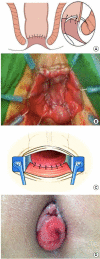
(A) Hand-sewn coloanal anastomosis with interrupted absorbable sutures between the colon, anal mucosa, and external sphincter. (B, C) Colonanal anastomosis result. (D) Prominent mucosal prolapse after intersphincteric resection causing wet anus. Adapted from Kim et al. [102] with permission of Springer.
Fig. 13.
Fig. 13.
Cadaveric dissection shows a clear cleavage plane between the internal and external anal sphincter.
Fig. 14.
Fig. 14.
Histologic studies (H&E stain, ×10) showed the internal anal sphincter (IAS, smooth muscle) and external anal sphincter muscle (EAS, skeletal muscle), along with the conjoined longitudinal muscle between them. Adapted from Lee et al. [114] according to the Creative Commons License.
Fig. 15.
Fig. 15.
Partial excision of levator ani muscle (LAM). DL, dentate line; IAS, internal anal sphincter; EAS, external anal sphincter; ISS, intersphincteric space; ISG, intersphincteric groove. Adapted from Yang et al. [115] with permission of Wiley.
Fig. 16.
Fig. 16.
Diagonal coloanal anastomosis for coloanal anastomosis for partial excision of the levator ani muscle. DL, dentate line; IAS, internal anal sphincter; EAS, external anal sphincter; ISS, intersphincteric space; ISG, intersphincteric groove.
Fig. 17.
Fig. 17.
Colonoscopy findings showed a diagonal anastomotic line in a patient who underwent partial excision of the levator ani muscle after preoperative chemoradiotherapy. A rectal exam can also confirm this oblique line.
Fig. 18.
Fig. 18.
Conventional defecography showed an acceptable anorectal angle oner after ileostomy closure in a patient who underwent partial excision of the levator ani muscle.
Fig. 19.
Fig. 19.
(A, B) Rectal magnetic resonance imaging showed tumor invaded to the levator ani muscle bilateral extensively to be a candidate for extralevator abdominoperineal resection. (C, D) However, it showed that the tumor invaded only partly to the levator ani muscle so that it can be a candidate for partial excision of the levator ani muscle. Adapted from Yang et al. [115] with permission of Wiley.
Fig. 20.
Fig. 20.
Comparison between preoperative (A) and postoperative (B) rectal magnetic resonance imaging showed an absence of right-side levator ani muscle plate. Adapted from Noh et al. [117] according to the Creative Commons License.
Fig. 21.
Fig. 21.
Pelvic magnetic resonance imaging shows enlarged right inguinal lymph node.
Fig. 22.
Fig. 22.
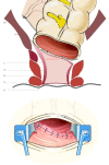
Two-stage Turnbull-Cutait pull-through coloanal anastomosis. (A) Exteriorization of the colon. (B) Colon fixation to the anal margin. (C) Hand-sewn coloanal anastomosis. Modified from Biondo et al. [32] with permission from JAMA.

Similar articles

Cited by

References

    1. Kim NK, Kim HS, Alessa M, Torky R. Optimal complete rectum mobilization focused on the anatomy of the pelvic fascia and autonomic nerves: 30 years of experience at Severance Hospital. Yonsei Med J. 2021;62:187–99. - PMC - PubMed
    1. Church JM, Raudkivi PJ, Hill GL. The surgical anatomy of the rectum: a review with particular relevance to the hazards of rectal mobilisation. Int J Colorectal Dis. 1987;2:158–66. - PubMed
    1. Kim NK. Anatomic basis of sharp pelvic dissection for curative resection of rectal cancer. Yonsei Med J. 2005;46:737–49. - PMC - PubMed
    1. Bogach J, Tsai S, Zbuk K, Wong R, Grubac V, Coates A, et al. Quality of preoperative pelvic computed tomography (CT) and magnetic resonance imaging (MRI) for rectal cancer in a region in Ontario: a retrospective population-based study. J Surg Oncol. 2018;117:1038–42. - PubMed
    1. Zhou Y, Shao W, Lu W. Diagnostic value of endorectal ultrasonography for rectal carcinoma: a meta-analysis. J Cancer Res Ther. 2014;10 Suppl:319–22. - PubMed

LinkOut - more resources