. 2021 Dec 28;7(1):122.
doi: 10.1038/s41421-021-00344-4.

Histone lysine methacrylation is a dynamic post-translational modification regulated by HAT1 and SIRT2

Affiliations

Histone lysine methacrylation is a dynamic post-translational modification regulated by HAT1 and SIRT2

Kyle Delaney et al. Cell Discov. .

Abstract

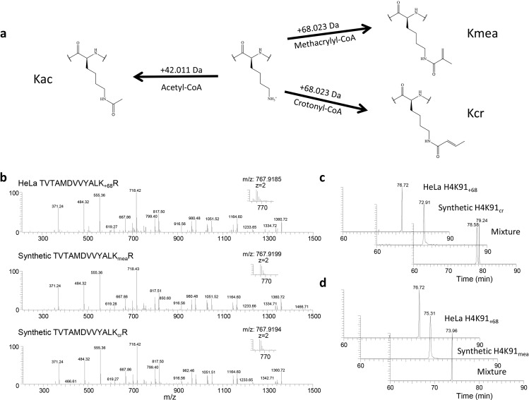
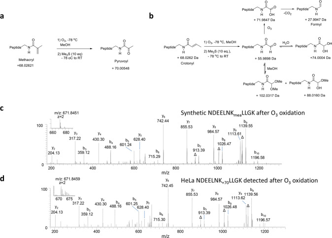
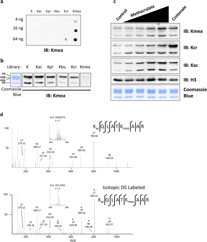
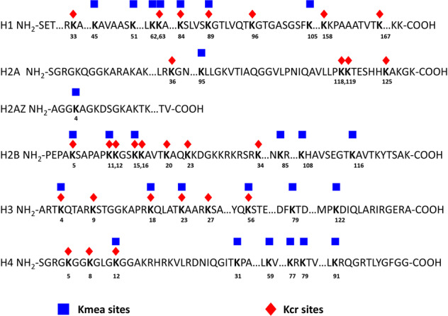
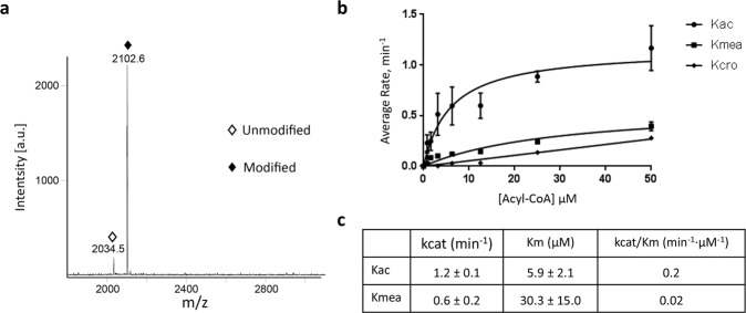
Histone lysine crotonylation is a posttranslational modification with demonstrated functions in transcriptional regulation. Here we report the discovery of a new type of histone posttranslational modification, lysine methacrylation (Kmea), corresponding to a structural isomer of crotonyllysine. We validate the identity of this modification using diverse chemical approaches and further confirm the occurrence of this type of histone mark by pan specific and site-specific anti-methacryllysine antibodies. In total, we identify 27 Kmea modified histone sites in HeLa cells using affinity enrichment with a pan Kmea antibody and mass spectrometry. Subsequent biochemical studies show that histone Kmea is a dynamic mark, which is controlled by HAT1 as a methacryltransferase and SIRT2 as a de-methacrylase. Altogether, these investigations uncover a new type of enzyme-catalyzed histone modification and suggest that methacrylyl-CoA generating metabolism is part of a growing number of epigenome-associated metabolic pathways.


Conflict of interest statement

Y.Z. is a founder, equity owner, board member, advisor to, and inventor on patents licensed to PTM Bio Inc. and Maponos Therapeutics Inc. The authors declare no competing interests.

Figures

Fig. 1
Fig. 1. Identification and verification of lysine methacrylation (Kmea).
a Lysine (center) is capable of being modified with multiple different PTMs depending on which acyl-CoA is used in the enzymatic reaction. The appropriate acyl-CoAs as well as the associated mass shift added to a lysine residue is indicated for acetyllysine (left), methacryllysine (Kmea, upper right), and crotonyllysine (Kcr, lower right). b MS/MS spectra of an exemplary HeLa histone peptide (top), synthetic H4K91ma peptide (middle), and synthetic H4K91cr peptide (bottom). c Extraction ion chromatograms of HeLa cells-derived histone peptide (TVTAMDVVYALK+68R) (top), synthetic H4K91cr peptide (middle), and a mixture of the two peptides (bottom). d Extraction ion chromatograms of HeLa cells-derived histone peptide (TVTAMDVVYALK+68R) (top), synthetic H4K91mea peptide (middle), and a mixture of the both peptides (bottom).
Fig. 2
Fig. 2. Verification of Kmea structure by ozone (O3) oxidation.
a, b Chemical reactions for ozonolysis of the double bonds in Kmea (a) and Kcr (b) peptides. c MS/MS of ozonolyzed peptides. The synthetic H2AK95mea peptide, NDEELNKmeaLLGK, was subjected to ozonolysis reaction. Its main reaction product, NDEELNK+70LLGK, was analyzed by HPLC/MS/MS analysis. d MS/MS of a NDEELNK +70LLGK peptide detected in O3 oxidized HeLa histone extract.
Fig. 3
Fig. 3. Methacrylate is a metabolic precursor for Kmea.
a Dot blot assay used to assess specificity of a pan anti-Kmea antibody. The Kmea peptide library consists of peptides with the sequence CXXXXXKmeaXXXXX where C is cysteine, X is a mixture of the 19 non-cysteine amino acids, and Kmea is methacryllysine. The control peptides libraries differ in modification status of the central lysine residue where the modifications are abbreviated as follows: K, unmodified lysine; Kac, acetyllysine; Kpr, propionyllysine; Kbu, butyryllysine; and Kcr, crotonyllysine. The amount of peptides spotted in each row of the membrane is indicated. b Western blotting was performed using a pan anti-Kmea antibody on acid extracted HeLa histones. The pan anti-Kmea antibody was pre-incubated with peptide libraries for 4 h prior to immunoblotting of the western blot membrane. The sequences and abbreviations of the peptide libraries are the same as those indicated for the dot blot in a. c Western blot results for histones extracted from HeLa cells. HeLa cells were treated for 24 h with 1, 3, 5, or 10 mM sodium methacrylate or 10 mM sodium crotonate. d MS/MS spectra of H3K23mea peptide from HeLa histones following treatment with either methacrylate (top) or d-7 methacrylate (bottom). Asterisks (*) indicate b and y ions with 5 Da mass shift consistent with incorporation of the isotopic metabolite.
Fig. 4
Fig. 4. Maps of the identified Kmea and Kcr on HeLa histones.
Schematic of the sequences of the 5 core histone proteins and 1 variant histone sequence (H2AZ) with sites of Kmea and Kcr identified in HeLa cells. The key for the modifications is as follows: Kmea, blue squares; Kcr, orange diamonds. Kmea sites include both those detected by IP-HPLC/MS/MS with pan Kmea antibody as well as sites that were validated by co-elution or ozonolysis in HeLa cells. Kcr sites were detected in our previous publication using HeLa cell histones.
Fig. 5
Fig. 5. HAT1 catalyzes Kmea addition in vitro.
a Synthetic peptides consisting of the first 20 amino acid residues of the H4 histone, H4 (1-20), were incubated with methacrylyl-CoA and recombinant HAT1 in vitro. MALDI-TOF analysis detected unmodified (open diamond) and methacrylated (solid diamond) peptides. b HAT1 was incubated with acetyl-CoA, methacrylyl- CoA, or crotonyl-CoA at varied concentrations and H4 (1-20) peptide substrates for 15 min, respectively. The enzymatic reaction was quantified using 7-diethylamino-3-(4’-maleimidylphenyl)-4-methylcoumarin (CPM) assay. c The reaction rate-acyl-CoA concentration was plotted with the Michaelis-Menten equation to get the kinetic constants Km and kcat for acetyl-CoA and methacrylyl-CoA substrates, respectively. The kcat/Km values were used to evaluate lysine acylation efficiency.
Fig. 6
Fig. 6. HAT1 regulates H4K5mea in cells.
a Close-up view of the HAT1-acetyl-CoA interaction site in the crystal structure of HAT1 in complex with acetyl-CoA and histone H4 peptide (PDB ID: 2P0W). HAT1 is shown in cartoon representation in gray, acetyl-CoA in green, and histone H4 in yellow. b Close-up view of the methacrylyl-CoA interaction site with HAT1 in a model generated by fitting the methacrylyl-CoA into the isobutyryl-CoA electron density map in the crystal structure of HAT1-isobutyryl-CoA (PDB ID: 6VO5) for illustration purposes. HAT1 is shown in cartoon representation in gray, methacrylyl-CoA in cyan and histone H4 in yellow. c HEK 293 T were transfected with empty pcDNA 3.1 vector or pcDNA 3.1 HAT1-Flag plasmid for 48 h before harvesting. Cell lysates were prepared using RIPA buffer and then analyzed by western blot. d HEK 293 T cells were transfected for 72 h with 25 nM of control or HAT1 siRNA. RIPA lysates were probed using indicated antibodies. e MEF HAT1−/− and MEF HAT1+/+ cells were grown in culture before harvesting lysates. RIPA lysates were assessed by western blot as indicated.
Fig. 7
Fig. 7. SIRT1 and SIRT2 remove methacryl from Kmea.
a Acid extracted HeLa histones were incubated with or without recombinant HDAC3 for 12 h at 37 °C. Samples were then analyzed by western blot. b HEK 293 T cells were treated for 24 h with either 10 mM sodium butyrate or 1 μM TSA. The core histones were prepared by acid extraction method and then subjected to western blot analysis. c, d Quantitation of deacylase activity of SIRT 1-7 for in vitro screen using either H3K18mea peptide (c) or H3K18ac peptide (d) is shown. The deacylase activity percentage was calculated by dividing the area of the H3K18 unmodified chromatogram peak over the sum of the H3K18 unmodified and modified chromatogram peaks. e H3K18mea synthetic peptides were incubated with SIRT1 or SIRT2 in presence of reaction buffer with or without its cofactor NAD + . Chromatograms of each sample condition are shown. Open diamonds indicate the H3K18 unmodified peptide peaks. Filled diamonds indicate the H3K18mea peptide peaks. f Acid-extracted HeLa histones were incubated with or without recombinant SIRT1 or SIRT2 for 12 h at 37 °C. Samples were then analyzed by western blot. g HEK 293 T cells were transfected with either control empty pcDNA 3.1 vector or pcDNA 3.1 SIRT2-Flag. Whole cell lysates were prepared 48 h post-transfection and then subjected to western blot analysis with the indicated antibodies.

References

    1. Diehl KL, Muir TW. Chromatin as a key consumer in the metabolite economy. Nat. Chem. Biol. 2020;16:620–629. - PMC - PubMed
    1. Lin H, Su X, He B. Protein lysine acylation and cysteine succination by intermediates of energy metabolism. ACS Chem. Biol. 2012;7:947–960. - PMC - PubMed
    1. Huang H, Sabari BR, Garcia BA, Allis CD, Zhao Y. SnapShot: histone modifications. Cell. 2014;159:458–458.e451. - PMC - PubMed
    1. Sabari BR, Zhang D, Allis CD, Zhao Y. Metabolic regulation of gene expression through histone acylations. Nat. Rev. Mol. Cell Biol. 2017;18:90–101. - PMC - PubMed
    1. Moellering RE, Cravatt BF. Functional lysine modification by an intrinsically reactive primary glycolytic metabolite. Science. 2013;341:549–553. - PMC - PubMed